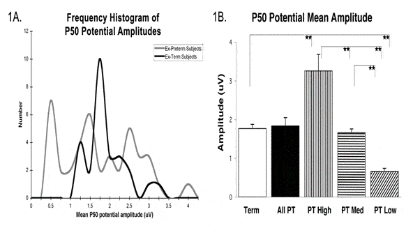
We needed to be able to measure the effects of illness, lesion, drugs, etc. on different levels of the neuraxis in humans. Therefore, we established the capacity for recording the midlatency auditory P50 evoked potential, whose amplitude is a measure of level of arousal and thus assesses brainstem-thalamus processes (5). The use of paired auditory stimuli allows us to measure habituation to repetitive stimulation, or sensory gating, a process disturbed in a number of diseases (5). We also developed the capacity to measure reaction time (RT) using a Psychomotor Vigilance Task (6) to derive the prototypical measure of attention and thalamocortical processes (7). Finally, we developed the capacity to measure frontal lobe blood flow using near infrared spectroscopy, which provides an economical indicator for the use of more expensive methods such as PET. We established a satellite facility for spinal reflex testing in spinal cord injury and other patient populations.
![P50 potential amplitude. A. The amplitude of the P50 potential is on the x-axis, and the frequency of occurrence is shown on the Y-axis. The distribution of P50 potential amplitudes in Ex-Term adolescents (dark line) was unimodal, whereas the distribution of P50 potential amplitudes in Ex-Preterm adolescents appeared to have three peaks (gray line). A test of Variance revealed a significant difference between the two distributions (p<0.005). A Mann-Whitney-U test of the two distributions revealed that they were statistically different (P<0.01). B. Mean amplitude of the P50 potential for all Ex-Term (open bar) and Ex-Preterm (filled bar) adolescents demonstrating no difference in amplitude when all Ex-Preterm subjects were pooled. The multimodal distribution of P50 amplitudes among Ex-Preterm subjects showed the presence of three peaks falling below the 33rd , between the 33rd and 67th, and above the 67th percentiles. Comparisons between High (vertical stripes), Medium (horizontal stripes) and Low (diagonal stripes) amplitude peaks of Ex-Preterm adolescents showed significant differences between peaks at **p<0.01](https://medicine.uams.edu/neurobiology/wp-content/uploads/sites/19/2022/09/Human-Electro-Core-image.gif)