• Skip to main content
  • Skip to main content
Choose which site to search.
University of Arkansas for Medical Sciences Logo University of Arkansas for Medical Sciences
Department of Neurobiology and Developmental Sciences: Center for Translational Neuroscience
  • UAMS Health
  • Jobs
  • Giving
  • About Us
    • Be a Part of the Cure
    • Contact Us
    • Distinguished Lecturer Series
    • Events
  • Core Facilities
    • Animal Electrophysiology Core
    • Human Brain Tissue Core
    • Human Electrophysiology Core
    • Imaging Core
    • Molecular Core
    • Telemedicine Core
    • Transcranial Magnetic Core
  • Faculty
  • Funding
  • Publications
  • Research
    • Research Highlights
      • Consciousness and Dreaming
      • Hyper-reflexia
      • Leptin
      • Modafinil
      • Preconscious Awareness
      • Premature Babies
      • Sleep-Wake Control
      • Telemedicine
      • Tinnitus
    • Brown Lab
    • Charlesworth Lab
    • Chen Lab
    • Dornhoffer Lab
    • Fantegrossi Lab
    • Garcia-Rill Lab
    • Hall Lab
    • Hayar Lab
    • Kaiser Lab
    • Kiser Lab
    • MacNicol Lab
    • Mancino Lab
    • Mennemeier Lab
    • Sheffer Lab
    • Skinner Lab
    • Yates Lab
  1. University of Arkansas for Medical Sciences
  2. College of Medicine
  3. Department of Neurobiology and Developmental Sciences
  4. Research Centers
  5. Center for Translational Neuroscience
  6. Research
  7. Skinner Lab

Skinner Lab

Robert D. Skinner, Ph.D.

skinnerrobertd@uams.edu

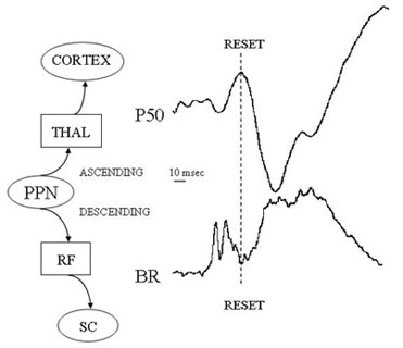
An intrinsic function of the cholinergic arm of the reticular activating system (RAS), the pedunculopontine nucleus (PPN), is its participation in “fight vs flight” responses such that alerting stimuli simultaneously activate higher thalamocortical (THAL, CORTEX) systems, as well as postural and locomotor systems (RF reticular formation, SC spinal cord), in order to enable an appropriate response. The P50 midlatency auditory-evoked potential appears to be an ascending manifestation of the cholinergic arm of the RAS in eliciting changes in arousal state in thalamo-cortical structures. Abnormalities in the manifestation of the P50 potential are present in disorders that include: 1) dysregulation of sleep-wake cycles; 2) abnormalities in reflex/postural, especially, startle and blink responses; and 3) malfunctions in flight vs flight responses. In general, the P50 potential appears to be upregulated (increased amplitude and/or decreased habituation) in disorders that are marked by upregulation of RAS outputs (hypervigilance), and downregulated in disorders characterized by decreased RAS outputs (hypovigilance). Many of the disorders discussed have a developmental etiology and a postpubertal age of onset, e.g. schizophrenia, anxiety disorders, depression. We have developed the rodent equivalent of the P50 potential, the rat P13 potential, which has the same characteristics of a) sleep state-dependence (present during waking and REM sleep, absent during slow wave sleep), b) mediated by cholinergic projections (blocked by muscarinic antagonist scopolamine) and c) rapid habituation (generated by multi-synaptic, low security “reticular” pathways). The figure below shows how the two waves occur during the inhibition of the electromyogram of the blink reflex (BR) or the startle response (SR).

decorative
UAMS College of Medicine LogoUAMS College of MedicineUniversity of Arkansas for Medical Sciences
Mailing Address: 4301 West Markham Street, Little Rock, AR 72205
Phone: (501) 686-7000
  • Facebook
  • Twitter
  • Instagram
  • YouTube
  • LinkedIn
  • Pinterest
  • Disclaimer
  • Terms of Use
  • Privacy

© 2023 University of Arkansas for Medical Sciences