Overview
The Histology and Bioimaging Core assists investigators in the Arkansas Children’s Nutrition Center with all aspects of routine histological and immunohistochemical procedures, and also helps investigators develop new procedures and special stains as needed. Daily activities including maintenance of equipment, project management and development and standardization of new techniques in the Core are overseen by full-time, certified histotechncian, Ms. Reneé Fox, HT(ASCP).
Visualizing the Smallest Tissues
From histochemical and immunohistochemical analysis, in situ hybridization, and imaging/microscopy of tissues and cells, The Arkansas Children’s Nutrition Center Histology and Bioimaging Core, has all the necessary facilities.

Precise and Consistent Sample Preparation
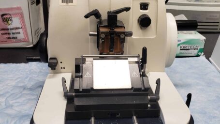
Leica RM2135 microtome is used for cutting paraffin embedded blocks

High-quality Tissue Processing
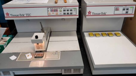
An automated Sakura Tissue-Tek VIP6 AI processor and a Tissue Tek Embedding platform for sample preparation

Efficiently Sectioned Tissue Samples
An Epredia Cryostar NX70 performs analyses on frozen sections

Microscopy
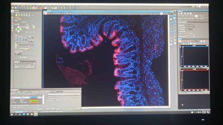
Several microscopes (Olympus BX50 and Nikon Eclipse Ti2) with histomorphometry, density scanning, 3-D reconstruction and rendering capabilities, and NIS Elements AR software
