Services
The services in the Core are divided into those that primarily serve to determine the size and composition of macromolecules and those that report on dynamics and equilibria. A description of services is provided below.
All users needing training before use of the instruments need to contact Dr. John Marecki (BioMed II, Room 433-2) to schedule time for training. Scheduling time for use of the instruments is through Outlook calendar system. Please contact Dr. Eric Enemark for authorization.
Differential Scanning Fluorometry (nanoDSF)
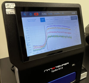 The Tycho instrument from NanoTemper Technologies measures changes in the intrinsic fluorescence of proteins during thermal unfolding, and this method is called nanoDSF. When the aromatic amino acids are exposed upon protein unfolding, both the intensity and the maximum wavelength of their fluorescence change. By measuring the ratio of the two emission wavelengths, Tycho can detect even a small shift of the intrinsic fluorescence for determining protein stability under various buffer and reaction conditions.
Microscale Thermophoresis (MST)
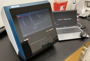 The MonolithX instrument from NanoTemper Technologies allows for the measurement of specific molecular interactions based on the principle of MST by monitoring microscopic temperature gradients that accompany changes in the solvation shell, charge state or size when two biomolecules encounter one another. Many types of molecules can be studied, including large macromolecules, small-molecules, ions, and even complex mixtures. MST allows rapid assessment of interactions with a wide range of affinities (i.e. pM to mM) using very little sample.
CMIC Usage for the MonolithX
Mass Photometry
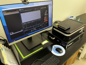 The TwoMP Mass photometry instrument from Refeyn, Inc. measures the molecular weight of biopolymers and multi-subunit complexes in solution at the level of single molecules. Similar to SEC-MALS, mass photometry can help inform structural approaches. Core users can obtain results faster and with less sample using mass photometry, and compared to SEC-MALS, the data analysis step is simpler.
Isothermal Titration Calorimetry (ITC)
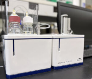 To perform quantitative, label-free analysis of macromolecular interactions, Core users have access to the Microcal PEAQ-ITC instrumentation and software from Malvern Panalytical. Estimates of equilibrium binding affinity made by ITC rely on precise measurement of the amount of heat released or taken up during each reaction. ITC is the only technique that directly measures the thermodynamic parameters of macromolecular interactions, allowing investigators to measure affinity, stoichiometry, and gain insight into the forces driving biomolecular interactions, such as those guiding PPIs or small-molecule inhibitor binding. Relatively little sample is required (~0.3-0.5 mL in the mM to mM range).
CMIC Usage for the ITC (Contact Dr. Amit Ketkar, CORE C)
Total Internal Reflection Fluorescence (TIRF) Microscopy
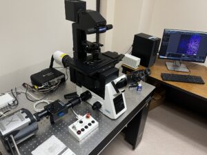 TIRF microscopy has exceptionally good signal-to-noise ratio and combines kinetic analysis with spatial information. The instrumentation housed in the Biomolecular Interactions Core allows RPLs and other Center members to design experiments that monitor fluorescent signal from single-molecules. Researchers can also perform experiments that measure the kinetics of protein localization, cytoskeletal changes, organelle movement, and cell-cell interactions.
Size-Exclusion Chromatography with Multi-Angle Light Scattering (SEC-MALS)
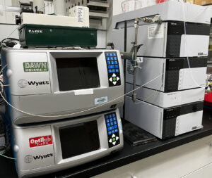 The SEC-MALS system allows investigators to determine the molar mass (M), size (rg), second virial coefficient (A2) and translational diffusion coefficient (Dt), which can be used to calculate the hydrodynamic radius (Rh) of macromolecules using first principles. A variety of biopolymers, including proteins, nucleic acids, carbohydrates, and liposomes can be measured in solution with SEC-MALS. Multiple oligomeric states can be separated and analyzed with a size range of around 200 Da through 1 GDa. The size range of SEC-MALS provides a slight advantage over mass photometry, but the two techniques are complementary in many ways. Acceptable signal-to-noise ratios can be achieved with less than a microgram of a monodisperse sample for proteins/molecules in the range of 100 kDa.
CMIC Usage for the SEC-MALS (Contact Dr. John Marecki)
Circular Dichroism (CD) Spectroscopy
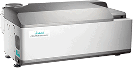 The application of CD spectroscopy to projects in the CMIC allows investigators to determine if molecules involved in oncogenic processes have altered folding patterns or changes in stability. CD is also useful for checking proper folding of recombinant proteins. Measurements typically require ~50-100 mL of solution containing analytes at a concentration in the mM range.
CMIC Usage of the Kendrick CD Spectrometer
Fluorescence Anisotropy
The measurement of changes in the fluorescence intensity of polarized light allows researchers to observe differences in the rotational diffusion of macromolecules that occur upon binding of another molecule or ligand. A wide variety of macromolecular interactions can be studied with this approach with analytes at low concentrations in relatively small volumes.
Resources for Fluorescence Anisotropy
More Information
For more information or to set up a consultation, please contact:


How to Acknowledge the CMIC
“Research reported in this [publication, release] was supported (in whole or in part) by the National Institute of General Medical Sciences of the National Institutes of Health under Award Number P20GM152281. The content is solely the responsibility of the authors and does not necessarily represent the official views of the National Institutes of Health.”
For abstracts, a short note “Supported by a grant from the NIH/NIGMS – 1P20GM152281” will suffice.