Blood’s Editor-in-Chief
We want to congratulate Dr. Storrie and his Lab for getting a journal cover from Blood’s Editor-in-Chief. They will be the cover of the May issue of Blood VTH

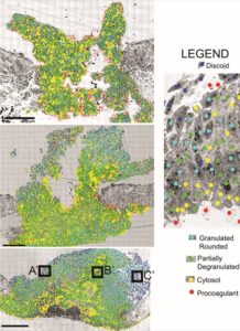
5-color, time series (top to bottom) of platelet activation state distribution in mouse jugular vein puncture wound thrombi. Mouse jugular vein was exposed and punctured with a 30-gauge needle, nominal diameter 310 microns. Samples were fixed at 1-, 5-, and 20-minutes post-puncture and prepared for transmission electron microscopy at 3.185 nm pixel size. 600-800 individual image frame were montaged together to give full thrombus cross sections and platelets in the resulting computer images were stratified/assigned to each of 5 activation states based on morphology and a-granule abundance. The distributions of the highest activation state platelets (red and yellow) were progressively buried from exposure to the intravascular circulation as the thrombi reorganized over time. The vessel wall is to the middle left and right of the thrombus and the intravascular portion of the image is up and the extravascular portion is down.
Cover Illustration (Below)
(The actual Journal cover will be available in a few days)