Assistant Professor
Office: Biomed 1, B323D
Lab: Biomed 1, B320
Phone: 501-686-5810
Email: afouda@uams.edu
LinkedIn profile
Download Dr. Fouda’s CV (PDF)
Education and Training
- B.Pharm – Cairo University School of Pharmacy, Egypt (2002 – 2007)
- Teaching/research assistant – Cairo University, Clinical Pharmacy Dept., Egypt (2007 – 2011)
- Ph.D. – University of Georgia, Clinical and Experimental Therapeutics program (2011 – 2015)
- Post-doctoral training – University of Georgia, Dr. Susan Fagan Stroke lab (2015 – 2016)
- Post-doctoral training – Augusta University, Dr. Ruth Caldwell Retinopathy lab (2016 – 2021)
Research Interests
Our lab studies central nervous system (CNS) injury mainly in the retina and brain. The lab employs different molecular/cell culture techniques, human tissue samples, and animal studies to model critical disease conditions in these two organs. Among the diseases under study are diabetic retinopathy, retinopathy of prematurity, traumatic optic neuropathy, traumatic brain injury, and stroke. Our studies aim to elucidate the underlying pathological mechanisms in these conditions and identify new therapies that can be translated from bench to bedside to help patients affected by these disease conditions.
We are interested in studying immune cells in CNS diseases with a special focus on the role played by myeloid cells (macrophages and microglia) as they respond to acute and chronic injury to the retina and brain. These cells closely interact with CNS neurons and vasculature and therefore play a major role in neurovascular injury and repair. Our goal is to gear the myeloid cells toward a reparative phenotype through modulation of different molecular pathways. The lab is funded by the National Eye Institute.
Fouda Lab Group
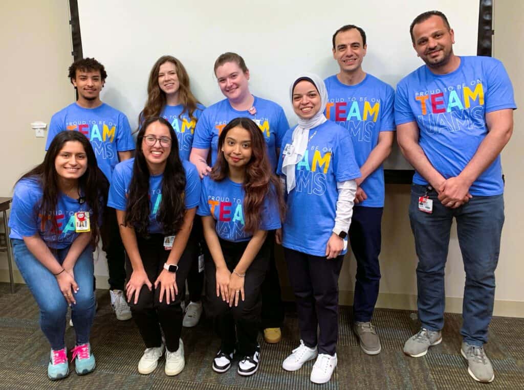
Meet Dr. Fouda’s Research Team

Fouda Lab News
Read the latest news from our lab!
Retina Imaging/Function Equipment

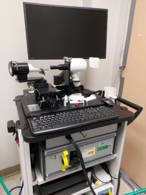
Retina imaging equipment (Bioptigen Spectral Domain Ophthalmic Imaging System (SDOIS; Bioptigen Envisu R2200))
The SDOIS is operated using proprietary Bioptigen software and features the DIVERS software for sophisticated measurements. It allows in vivo acquiring of optical coherence tomography (OCT) images of cornea tissue or retinal layers. OCT is a non-destructive alternative to histology thus enabling longitudinal studies with 10x fewer animals sacrificed. Diver software provides automatic segmentation of retinal layers and precise histologic analysis to avoid experimenter bias. It can be used to characterize the progression of disease in the same animal.

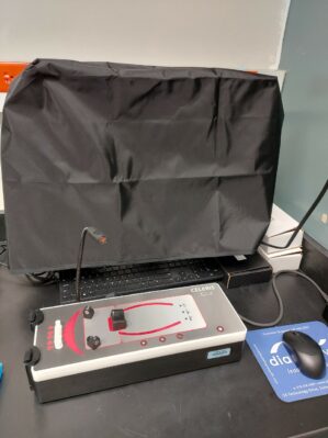
Celeris electroretinography (ERG)
This is a fully-integrated ERG system to measure the rodent retina function and assess the disease progression and effect of treatment in preclinical retinopathy research studies. The system enables high throughput and reproducible results, best suited for routine ERGs – such as scotopic (dim) and photopic (light) ERGs. It is also capable of performing pattern ERG (or PERG).
Research Support
Ongoing
- (6/2024 – 5/2025) UAMS Hornick award for stroke research
- (07/2021 – 06/2024) R00 EY029373 NIH/NEI
Role: PI Title: Role of Arginase 1 in Retinal Ischemia-Reperfusion Injury - 07/2021 – UAMS COM Startup funds
Role: PI
Completed
- K99 EY029373-01A1 NIH/NEI (09/2019 – 06/2021)
Role: PI Title: Role of Arginase 1 in Retinal Ischemia-Reperfusion Injury - 18POST34060036 AHA Postdoctoral Fellowship (07/01/2018 – 06/31/2020)
Role: PI Title: Arginase in retinal injury - University-Wide Graduate School Assistantship, UGA 8/2011 – 5/2013
