Initial Contact
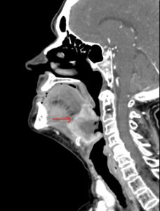
In 2013, a 68-year-old woman was diagnosed with squamous cell carcinoma at the base of the tongue. She underwent surgery consisting of removal of the mass and a left modified neck mode dissection, followed by chemotherapy and radiation. In June 2023, a surveillance CT scan showed the cancer had returned.
Assessment
The scan revealed a large necrotic tongue mass measuring almost 5 cm with invasion into the intrinsic muscles of tongue, epiglottis and paraglottic region. A PET-CT evaluation confirmed a soft-tissue mass along the base of the tongue, with extension, as noted above.
Mauricio Moreno, M.D., a surgeon with the UAMS Department of Otolaryngology-Head and Neck Surgery, performed a total glossectomy, total pharyngectomy, left hemithyroidectomy, neck nodes dissection and reconstruction with anterolateral thigh tissue transfer. However, post-op pathology indicated invasive squamous cell cancer measuring almost 9 cm, which was positive for perineural and extensive lymphovascular invasion, staging it as pT4a N0M0.
The head and neck surgery team met with medical oncologists, radiation oncologists and radiologists at UAMS, and adjuvant radiation treatment with concurrent chemotherapy was recommended.
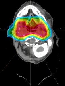
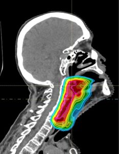
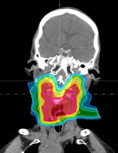
Given the prior history of radiation treatment, Santanu Samanta, M.D., an assistant professor in the UAMS Department of Radiation Oncology, recommended proton therapy. This would precisely target radiation at the post-op surgical site to prevent recurrence of the cancer while significantly reducing radiation to the surrounding structures: the spinal cord, brain stem, esophagus, brain, oral cavity and nasal cavity.
Procedure
Treatment occurred at the Proton Center of Arkansas at UAMS, which opened Sept. 27, 2023, as a collaboration between UAMS, Arkansas Children’s, Baptist Health and Proton International. It is the only proton center in Arkansas.
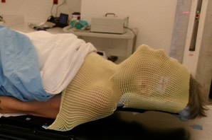
On an initial visit, the patient lay face-up while a mask was made for her face and neck that she could wear during subsequent visits. Then a CT scan was performed to allow a customized radiation plan to be developed, with special attention given to how the radiation beams would come from different directions to precisely target the post-op site while minimizing toxicity to the critical structures. Quality assurance checks followed to ensure the plan’s safety and effectiveness.
The radiation was delivered during subsequent visits lasting 30 minutes a day, five days a week, for a total of 30 treatments over six weeks. During each session, the patient lay on her back on a special couch, wearing the mask, and a low-energy CT scan ensured that she remained in the correct position while the proton therapy was delivered.
Proton therapy works by delivering energy precisely to the target to which it is directed, using protons generated from a cyclotron, a type of particle accelerator. The energy carried by the proton particles stop at the target, damaging the cancer cells by destroying the DNA in the nucleus of the cancer cells. But because the proton particles stop at a specific depth, no energy or radiation escapes beyond the target.
Unlike standard radiation with X-rays, there is no exit dose, and this makes proton therapy more precise than standard radiation.
Follow-up
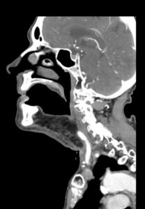
After completion of the treatment, the patient returned to the clinic about two months later. She underwent a CT scan that showed no evidence of recurrence. She had fully recovered from the side effects of radiation treatment, which included some soreness, dry mouth and sticky saliva, all of which are much less intense with proton therapy.
Discussion
The patient, who had a recurrence of cancer in the oral cavity, had undergone extensive surgery 10 years earlier, and based on the pathology report, had a high chance of recurrence without any adjuvant radiation.
It is important to note that a second course of radiation could be very challenging for someone who previously had a full course of radiation, because standard radiation treatment could deliver unnecessary and potentially harmful extra radiation to the spinal cord, brainstem, oral cavity and nasal cavity.
Hence, proton therapy is essential in this type of situation. Proton therapy provides a precise radiation dose to the post-op surgical site to reduce the chance of recurrence. It also minimizes radiation to the nearby structures — notably, the spinal cord, brain scan, nasal cavity, oral cavity, esophagus and lung.
This unique technology is for patients who need a repeat course of radiation treatments, as well as for pediatric patients for whom radiation treatment is absolutely indicated.