• Skip to main content
  • Skip to main content
Choose which site to search.
University of Arkansas for Medical Sciences Logo University of Arkansas for Medical Sciences
College of Medicine: Department of Radiology
  • UAMS Health
  • Jobs
  • Giving
  • About Us
    • News
    • Radiology Alliance for Community & Engagement
    • Giving
    • Contact Us
    • Where We Work
    • Faculty Recruitment
  • Faculty
  • Divisions
    • Neurointerventional Radiology
      • About Us
    • Interventional Radiology
    • Pediatric Radiology
  • Residency Programs
    • Diagnostic Radiology Residency Program
      • Meet Our Residents
      • Your Education
        • Pediatric Radiology On Call Survival Guide
        • Internship Year
        • Boot Camp
        • R1 Education
        • Conferences
        • Clinical Rotations
        • On Call Coverage
        • Mentoring
        • Board Prep
        • Moonlighting
      • Facilities
      • Living in Little Rock
      • Fellowship Placements
      • Wellness
      • Salary and Benefits
    • Interventional Radiology Integrated Residency
      • Guidelines for Selection
      • Clinical Sites
      • Training
      • Faculty
      • Meet Our Residents
      • Program Benefits
    • Interventional Radiology Independent Residency Program
      • Guidelines for Selection
      • Clinical Rotation Sites
      • Block Diagram
      • Training
      • Meet Our Faculty
      • Meet our Resident
      • Benefits
    • Early Specialization in Interventional Radiology Program
    • Newsletter Archive
    • Nuclear Medicine Training Program
    • Application Process
      • Frequently Asked Questions
    • Awards
    • Resident Scrapbook
  • Fellowships
    • Body MRI
    • Breast Imaging
    • Neuroradiology
    • Pediatric Radiology
  • Medical Students
    • Interventional Radiology Interest Group
    • Radiology Interest Group
    • Senior Electives
    • Summer in Radiology
    • The Dr. and Mrs. W.C. Culp Research Student Grant
    • M3s Secure
    • M4s Secure
    • Radiology Technology
  • Research
    • 2026 UAMS Department of Radiology Faculty Research Seed Grant Award
    • Annual Conferences
    • Ongoing Research
    • Presentations and Posters
    • Recent Publications
    • Resident Research
    • Research Help
    • Howard J.  Barnhard, MD, Radiology Research Symposium
  1. University of Arkansas for Medical Sciences
  2. College of Medicine
  3. Department of Radiology
  4. Pneumothorax, Pneumomediastinum – Air Leaks In The ICU

Pneumothorax, Pneumomediastinum – Air Leaks In The ICU

Last modified: October 8, 2022
You are here:
  • KB Home
  • ICU
  • Pneumothorax, Pneumomediastinum – Air Leaks In The ICU

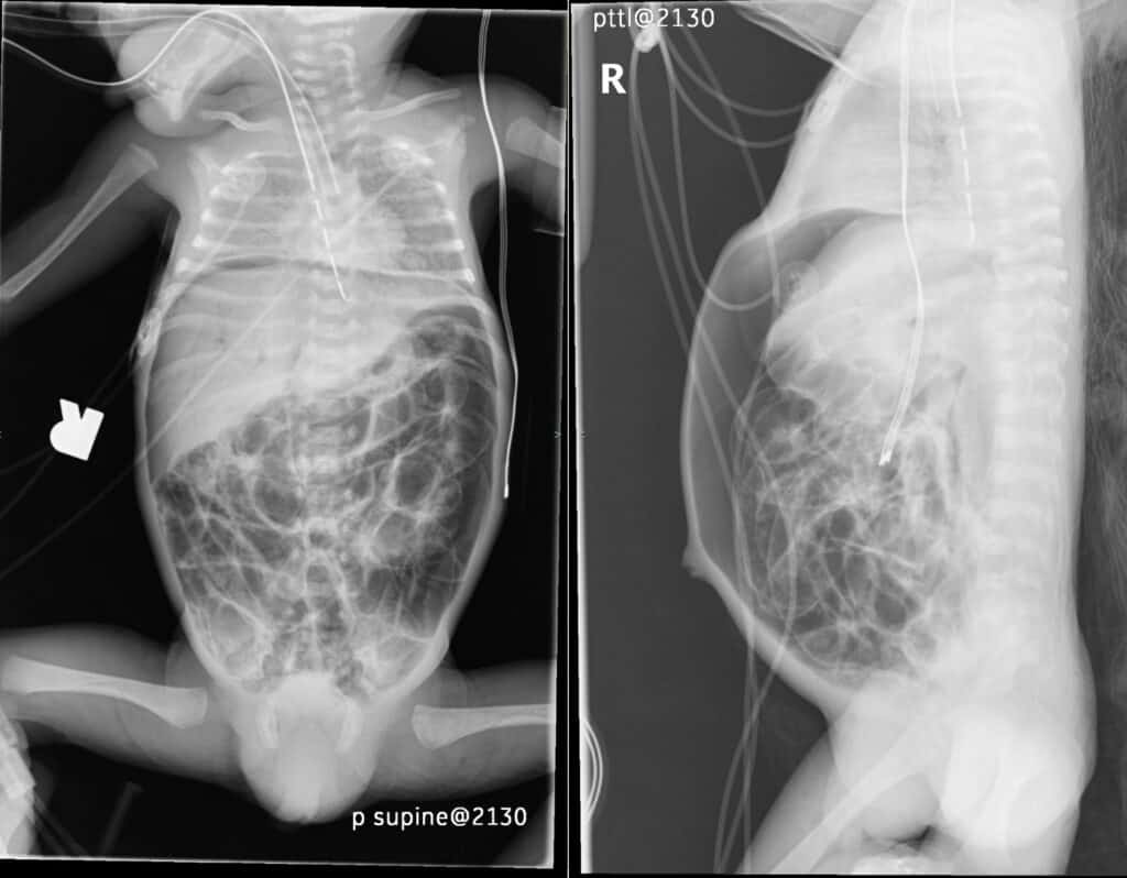
Most newborns (commonly preterms) needing additional respiratory support are placed on mechanical ventilation in the intensive care unit, which increases the risk of air dissecting through the alveolar spaces into the pulmonary interstitium with eventual rupture of air into the pleural cavity (4, 6).Useful signs in the diagnosis of small pneumothoraces include the deep sulcus sign (sharp and lucent costophrenic angle on the supine radiograph), medial stripe sign (increased lucency at the interface of the mediastinum and medial lung in case of a medial pneumothorax) and a hyperlucent hemithorax (4).

Axial CT Chest (non contrast) - small pneumothorax
Axial non-contrast chest CT through the lung apices demonstrates a small right apical pneumothorax with bubbly appearance of the anterior mediastinum reflecting pneumomediastinum.

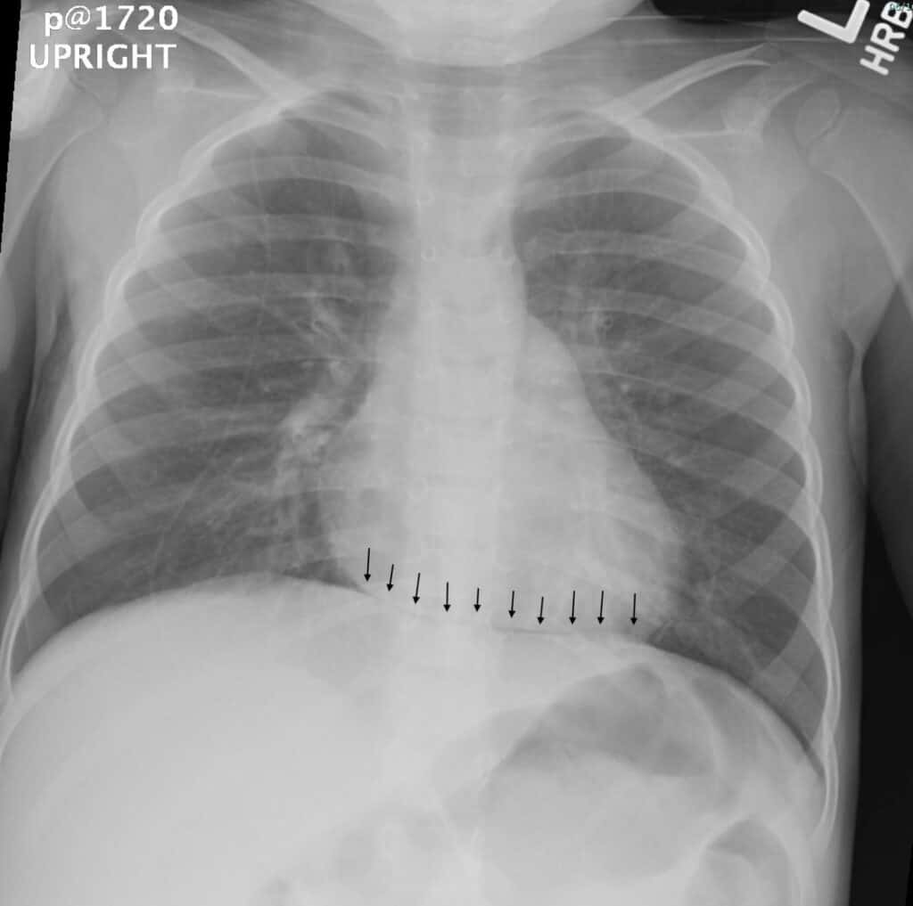
Pneumomediastinum can also accompany pneumothorax as a complication of positive pressure ventilation and manifests as accumulation of air in the mediastinum pushing the normal thymus upward and laterally (5, 6) which results in the characteristic angel wing sign.

Chest X-ray - Continuous Diaphragm Sign
Frontal chest radiograph of this infant’s chest demonstrates increased lucency in the subpulmonic recesses of both lungs which tracks along the inferior margin of the cardiac silhouette, crossing the midline into the contralateral side giving rise to the “continuous diaphragm” sign (black arrows).
Frontal and Cross Table X-Rays - Pneumatosis Intestinalis
Frontal and cross-table lateral radiographs of this infant’s chest and abdomen demonstrate extensive free air outlining the hepatic contour, prominent portal venous gas as well as bubbly lucency within the bowel indicating pneumatosis. The large amount of free air within the peritoneum is outlining multiple loops of bowel. This sign is called the Rigler’s sign. Football sign is another radiographic sign used to describe free air contained within the peritoneum and is well demonstrated on the lateral radiograph.

Selected References

  1. Bickle, I., & Shetty, A. (2020). Neonatal pneumothorax | Radiology Reference Article | Radiopaedia.org. Retrieved 13 September 2020, from https://radiopaedia.org/articles/neonatal-pneumothorax?lang=us
  2. Akin, K., Nevzat Cizmeci, M., Kenan Kanburoglu, M., Zulfikar Akelma, A., & Mansur Tatli, M. (2013). Angel Wing Sign in a Neonate with Pneumomediastinum. The Journal of Pediatrics, 163, 296. https://doi.org/10.1016/j.jpeds.2013.02.031de
  3. Lange, C. (2011). Radiology in paediatric non-traumatic thoracic emergencies. Insights into Imaging, 2(5), 585–598. https://doi.org/10.1007/s13244-011-0113-4

Was this article helpful?
Like 2 Dislike 0
UAMS College of Medicine LogoUAMS College of MedicineUniversity of Arkansas for Medical Sciences
Mailing Address: 4301 West Markham Street, Little Rock, AR 72205
Phone: (501) 686-7000
  • Facebook
  • X
  • Instagram
  • YouTube
  • LinkedIn
  • Pinterest
  • Disclaimer
  • Terms of Use
  • Privacy Statement
  • Legal Notices

© 2026 University of Arkansas for Medical Sciences