• Skip to main content
  • Skip to main content
Choose which site to search.
University of Arkansas for Medical Sciences Logo University of Arkansas for Medical Sciences
College of Medicine: Department of Radiology
  • UAMS Health
  • Jobs
  • Giving
  • About Us
    • News
    • Radiology Alliance for Community & Engagement
    • Giving
    • Contact Us
    • Where We Work
    • Faculty Recruitment
  • Faculty
  • Divisions
    • Neurointerventional Radiology
      • About Us
    • Interventional Radiology
    • Pediatric Radiology
  • Residency Programs
    • Diagnostic Radiology Residency Program
      • Meet Our Residents
      • Your Education
        • Pediatric Radiology On Call Survival Guide
        • Internship Year
        • Boot Camp
        • R1 Education
        • Conferences
        • Clinical Rotations
        • On Call Coverage
        • Mentoring
        • Board Prep
        • Moonlighting
      • Facilities
      • Living in Little Rock
      • Fellowship Placements
      • Wellness
      • Salary and Benefits
    • Interventional Radiology Integrated Residency
      • Guidelines for Selection
      • Clinical Sites
      • Training
      • Faculty
      • Meet Our Residents
      • Program Benefits
    • Interventional Radiology Independent Residency Program
      • Guidelines for Selection
      • Clinical Rotation Sites
      • Block Diagram
      • Training
      • Meet Our Faculty
      • Meet our Resident
      • Benefits
    • Early Specialization in Interventional Radiology Program
    • Nuclear Medicine Training Program
    • Application Process
      • Frequently Asked Questions
    • Awards
    • Resident Scrapbook
  • Fellowships
    • Body MRI
    • Breast Imaging
    • Neuroradiology
    • Pediatric Radiology
  • Medical Students
    • Interventional Radiology Interest Group
    • Radiology Interest Group
    • Senior Electives
    • Summer in Radiology
    • The Dr. and Mrs. W.C. Culp Research Student Grant
    • M3s Secure
    • M4s Secure
    • Radiology Technology
  • Research
    • Annual Conferences
    • Ongoing Research
    • Presentations and Posters
    • Recent Publications
    • Resident Research
    • Research Help
    • Radiology Research Symposium
  1. University of Arkansas for Medical Sciences
  2. College of Medicine
  3. Department of Radiology
  4. Non Accidental Head Trauma

Non Accidental Head Trauma

Last modified: October 8, 2022
You are here:
  • KB Home
  • Neuro
  • Non Accidental Head Trauma

What is it?

Approximately 1 in 3 children subjected to non-accidental trauma experience abusive head trauma, which unfortunately remains a major cause of morbidity and mortality in the abused pediatric population with mortality rates of more than 15% (6, 7). 

How does it happen?

Strangulation results in the classic hypoxic-ischemic injury pattern, which is detailed below. Also note, bimanual assault usually produces bilateral injuries, which may be the first sign of strangling (7).  

What do I need to know?

There are two main categories of neurological assault, shaking injuries (leading to subdural and retinal hemorrhages), and direct impact trauma causing coup/contrecoup injuries and skull fractures. Commonly, impact type injuries can cause depressed skull fractures, cortical contusions and extradural hemorrhage. Most common presentations of non-accidental trauma include – in order – interhemispheric subdural hematomas, cerebral edema and subarachnoid hemorrhage followed by infarct/ischemia (5).  

Axial FLAIR image demonstrates hyperintense subdural hematomas predominantly located in the bilateral frontoparietal convexity. Note that the most sensitive standard sequence is FLAIR as the appearance of a hematoma varies with the biochemical state of hemoglobin which is dependent on the age of the hematoma resulting in varying intensity on the T1 and T2 MR sequences. 

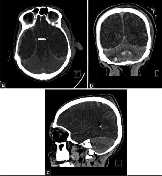
The classic ‘white cerebellum sign’ documented in pediatric victims of assault with irreversible hypoxic brain damage is uncommonly seen and denotes a poor prognosis (8). It is believed that the brainstem, cerebellum and thalamus may be selectively spared in early ischemia due to preservation of posterior circulation by autoregulation (7). 

Computed tomography scans in (a) Axial, (b) Coronal, and (c) Sagittal sections showing with right-sided acute subdural hematoma, bilateral hypodense cerebral hemispheres, and brain stem, complete cisternal effacement, and patchy contusions. The
Computed tomography scans in (a) Axial, (b) Coronal, and (c) Sagittal sections showing with right-sided acute subdural hematoma, bilateral hypodense cerebral hemispheres, and brain stem, complete cisternal effacement, and patchy contusions. The cerebellum is hyperdense to the rest of the brain.  Image courtesy: Krishnan P, Chowdhury SR. “White cerebellum” sign – A dark prognosticator. J Neurosci Rural Pract. 2014;5(4):433. doi:10.4103/0976-3147.140015 

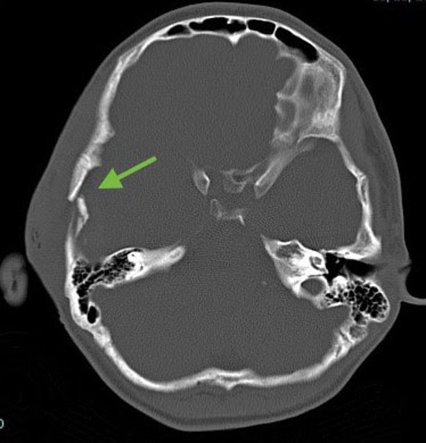
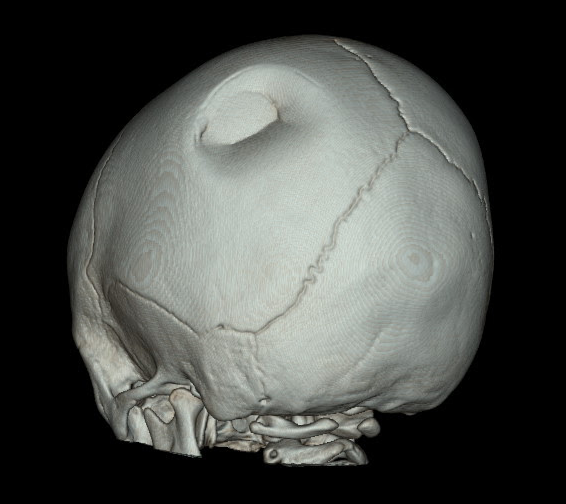
Depressed skull fractures are frequently associated with underlying intracranial injury. In our institution, all pediatric low dose trauma CT scans of the head include axial, sagittal and coronal reformations as well as 3D reconstructions to distinguish true fractures from normal sutures.  

axial head ct image showing skull fracture
Axial bone window reconstructions. Arrow points to depressed slightly comminuted and mildly displaced right temporal bone skull fracture. 

axial head CT image
Same slice on the axial blood window and soft tissue reconstruction software demonstrates a small lenticular and extra-axial collection compatible with acute traumatic subdural hematoma secondary to the overlying associated skull fracture (classic presentation). 

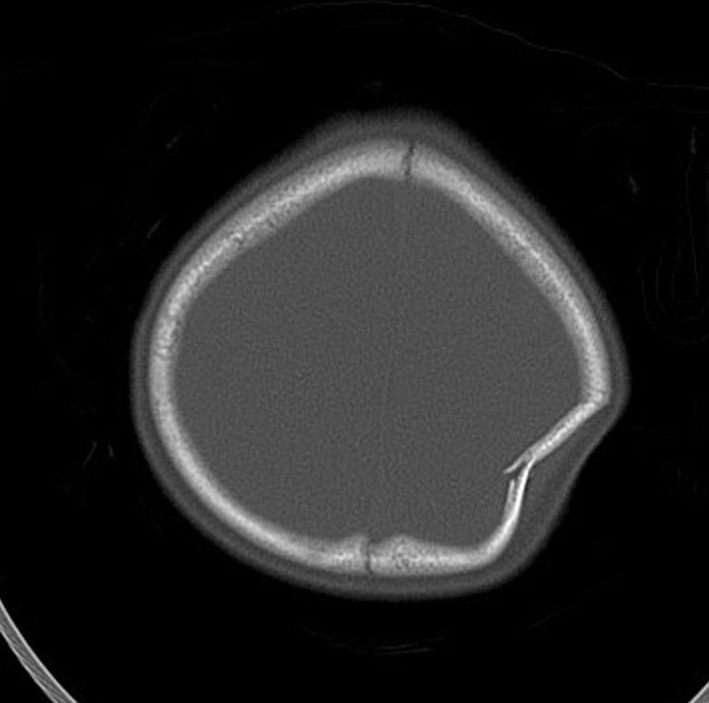
Ping pong fractures are specific skull fractures seen in neonates with soft and resilient calvaria which are able to indent without sustaining an actual break in the bone. This is similar to the mechanism for greenstick fractures in the long bones. These fractures are rarely associated with intracranial injury (9).  

axial CT in bone windows showing parietal bone fracture
Depressed left parietal bone fracture with complete cortical break is demonstrated to best advantage of this axial bone reconstruction image of the skull. 
  
3D skull reconstruction
3D reconstruction of the patient’s skull demonstrates the classic “ping pong” indentation in the left parietal bone. 

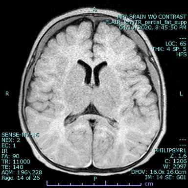
Although CT is usually sufficient to diagnose non accidental head injuries in children, MRI can be obtained for additional signs of injury and confirming evidence of repeated injuries (2).  

What do I need to do?

Call the emergency department to discuss the findings with the ER physician and document it on the preliminary report.  


Selected References

2. Huang, B. Y., & Castillo, M. (2008). Hypoxic-Ischemic Brain Injury: Imaging Findings from Birth to Adulthood. Radiographics, 28, 417–419. https://doi.org/10.1148/rg.282075066 

5. Ashwal, S., Wycliffe, N. D., & Holshouser, B. A. (2011). Advanced neuroimaging in children with nonaccidental trauma. Developmental Neuroscience, 32(5–6), 343–360. https://doi.org/10.1159/000316801 

6. Rolfes, M., Julie, G., Brucker, J., & Kalina, P. (2019). Article – Neuroimaging of pediatric abusive head trauma. Applied Radiology, 3, 30–38. https://www.appliedradiology.com/communities/Pediatric-Imaging/neuroimaging-of-pediatric-abusive-head-trauma 

7. Hsieh, K. L.-C., Zimmerman, R. A., Kao, H. W., & Chen, C.-Y. (2015). Revisiting Neuroimaging of Abusive Head Trauma in Infants and Young Children. American Journal of Roentgenology, 204(5), 944–952. https://doi.org/10.2214/AJR.14.13228 

8. An, M., Abam R, & Mp, O. (2019). White Cerebellum Sign-An Ominous Radiological Imaging Finding: A Case Report and Review of the Literature. Biomedical Journal of Scientific and Technical Research, 15(1), 11159–11161. https://doi.org/10.26717/BJSTR.2019.15.002659 

9. Zia, Z., Morris, A.-M., & Paw, R. (2007). Ping-pong fracture. Emergency Medicine Journal : EMJ, 24(10), 731. https://doi.org/10.1136/emj.2006.043570 

Was this article helpful?
Like 4 Dislike 0
UAMS College of Medicine LogoUAMS College of MedicineUniversity of Arkansas for Medical Sciences
Mailing Address: 4301 West Markham Street, Little Rock, AR 72205
Phone: (501) 686-7000
  • Facebook
  • X
  • Instagram
  • YouTube
  • LinkedIn
  • Pinterest
  • Disclaimer
  • Terms of Use
  • Privacy Statement

© 2025 University of Arkansas for Medical Sciences