• Skip to main content
  • Skip to main content
Choose which site to search.
University of Arkansas for Medical Sciences Logo University of Arkansas for Medical Sciences
College of Medicine: Department of Radiology
  • UAMS Health
  • Jobs
  • Giving
  • About Us
    • News
    • Radiology Alliance for Community & Engagement
    • Giving
    • Contact Us
    • Where We Work
    • Faculty Recruitment
  • Faculty
  • Divisions
    • Neurointerventional Radiology
      • About Us
    • Interventional Radiology
    • Pediatric Radiology
  • Residency Programs
    • Diagnostic Radiology Residency Program
      • Meet Our Residents
      • Your Education
        • Pediatric Radiology On Call Survival Guide
        • Internship Year
        • Boot Camp
        • R1 Education
        • Conferences
        • Clinical Rotations
        • On Call Coverage
        • Mentoring
        • Board Prep
        • Moonlighting
      • Facilities
      • Living in Little Rock
      • Fellowship Placements
      • Wellness
      • Salary and Benefits
    • Interventional Radiology Integrated Residency
      • Guidelines for Selection
      • Clinical Sites
      • Training
      • Faculty
      • Meet Our Residents
      • Program Benefits
    • Interventional Radiology Independent Residency Program
      • Guidelines for Selection
      • Clinical Rotation Sites
      • Block Diagram
      • Training
      • Meet Our Faculty
      • Meet our Resident
      • Benefits
    • Early Specialization in Interventional Radiology Program
    • Newsletter Archive
    • Nuclear Medicine Training Program
    • Application Process
      • Frequently Asked Questions
    • Awards
    • Resident Scrapbook
  • Fellowships
    • Body MRI
    • Breast Imaging
    • Neuroradiology
    • Pediatric Radiology
  • Medical Students
    • Interventional Radiology Interest Group
    • Radiology Interest Group
    • Senior Electives
    • Summer in Radiology
    • The Dr. and Mrs. W.C. Culp Research Student Grant
    • M3s Secure
    • M4s Secure
    • Radiology Technology
  • Research
    • 2026 UAMS Department of Radiology Faculty Research Seed Grant Award
    • Annual Conferences
    • Ongoing Research
    • Presentations and Posters
    • Recent Publications
    • Resident Research
    • Research Help
    • Howard J.  Barnhard, MD, Radiology Research Symposium
  1. University of Arkansas for Medical Sciences
  2. College of Medicine
  3. Department of Radiology
  4. Nonaccidental Trauma

Nonaccidental Trauma

Last modified: October 8, 2022
You are here:
  • KB Home
  • Musculoskeletal
  • Nonaccidental Trauma

What is it?

Basically, child abuse. Radiologists can often be the first to suspect non accidental injuries as these cases are difficult to recognize clinically. Overt clinical signs and symptoms of physical abuse may be subtle, caretakers frequently provide an inconsistent and unreliable history and presentation can be mistaken for other common pediatric diagnosis.

What do I need to know?

Musculoskeletal injury may be the chief complaint in a child subjected to physical abuse and therefore, it is critical to have a high suspicion of index for non-accidental trauma. Skeletal surveys are usually performed in all cases of suspected abuse.

Pathognomonic MSK (and neuro) findings to be aware of on call include:

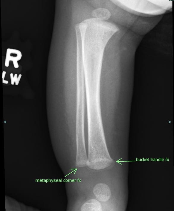
  • Characteristic corner/bucket handle fractures (Metaphyseal corner fractures) Mechanism: twisting injury or torsion under traction with consequent separation of the corner piece of the metaphysis from the remainder of the bone – More a shaking type injury with limbs flailing.
Frontal radiograph of the right distal lower extremity
Frontal radiograph of the right distal lower extremity. Note bucket-handle tear and metaphyseal corner fracture in the distal tibia and fibula, respectively.
  • Fractures in hard to break places (scapula, spinous process, sternum)
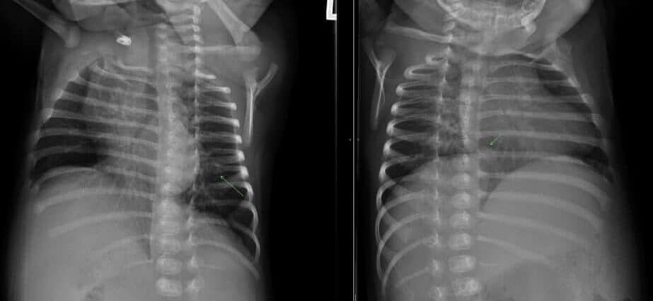
  • Rib fractures in multiple stages of healing (specifically, posterior rib)- Bilateral fractures
Oblique Chest Radiograph - Rib fractures suspicious of non accidental trauma
Oblique radiographs of the ribs demonstrating callus formation along the posterior body of the left 8th rib. Careful observation for posterior rib fractures (which may be of varying ages in multiple ribs) is highly suspicious for non accidental trauma.
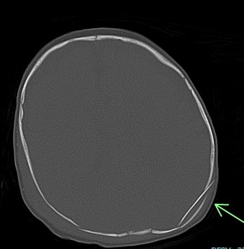
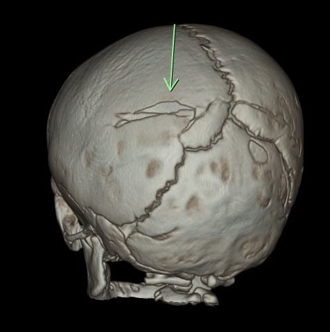
  • Skull fractures (best to see them on 3D bone recons)
    • Non parietal skull fractures
    • Crossing suture lines
    • Multiple bones
    • Depressed skull fracture (signature fracture)
Axial CT Brain - depressed ping pong like fracture
Axial CT images of the brain in bone windows with an arrow pointing to depressed, ping-pong like fracture of the left posterior parietal bone. Note minimally displaced fracture of the occipital and parietal bones on the right.
3D reconstructions of calvarium - comminuted & depressed skull fracture
Three-dimensional reconstructions of the calvarium in the same patient can better demonstrate the comminuted and depressed left posterior parietal bone fracture.
3D skull reconstruction - comminuted bone fracture
3D images of the skull highlight the comminuted right parietal bone fracture (arrow) in a different patient.
  • Spiral humeral and femoral fractures
Right Lower Extremity Radiograph - Spiral Fracture
Spiral fracture of the right mid femoral diaphyseal shaft
Lateral radiograph of Right proximal lower extremity - widely displaced spiral fracture
Lateral radiograph of the right proximal lower extremity in a different patient demonstrating a widely displaced spiral fracture of the humeral diaphysis with associated significant intramuscular hematoma.
  • Extra-axial hemorrhage (commonly subdural, may be epidural if associated with overlying skull fracture)
Axial noncontract CT head - traumatic subdural hemorrhage evaluation
Axial noncontrast CT of the head demonstrates extra-axial CSF density tracking along the bilateral cerebral convexities, compatible with traumatic subdural hemorrhage.
Brain MRI with T2 hyperintensities - traumatic subdural hemorrhage evaluation
Note bilateral prominence of the extra-axial space with T2 hyperintense fluid tracking along the cerebral convexities in keeping with traumatic subdural hemorrhage.
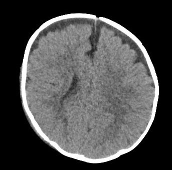
  • Coup, contrecoup injury associated with parenchymal contusions, commonly in the temporal and frontal lobes adjacent to the petrous bone and posterior to the greater wings of sphenoid as well as superior to the orbital roof, planum sphenoidale and cribriform plate in the frontal lobe (use sagittal and coronal recons if available)
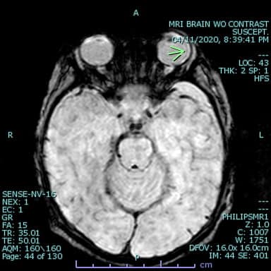
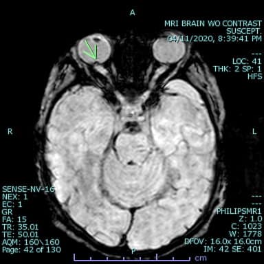
  • Retinal hemorrhage (hard to see but can be seen in susceptibility images on MR as areas of signal loss within the vitreous)In case of suspected physical abuse in a child ≤24 months of age and/or a child presenting with one of the following: neurologic signs, apnea, complex skull fracture, other fractures or injuries highly suspicious for child abuse, suspected thoracic or abdominopelvic injuries, initial imaging evaluation includes a complete skeletal survey per the ACR appropriateness criteria revised in 2016.
MRI Axial susceptibility weighted images of Orbits
MRI Axial susceptibility weighted images of Orbits (A)
MRI Axial susceptibility weighted images of Orbits
Axial susceptibility weighted images through the orbits demonstrate blooming artifact in the posterior aspect of the right globe in the region of the optic disc concerning for retinal hemorrhage (image B, arrow).

What do I need to do?

If you see even one of the above listed findings, call the emergency department and discuss the findings with the ER physician and document it on the preliminary report.


Was this article helpful?
Like 16 Dislike 0
UAMS College of Medicine LogoUAMS College of MedicineUniversity of Arkansas for Medical Sciences
Mailing Address: 4301 West Markham Street, Little Rock, AR 72205
Phone: (501) 686-7000
  • Facebook
  • X
  • Instagram
  • YouTube
  • LinkedIn
  • Pinterest
  • Disclaimer
  • Terms of Use
  • Privacy Statement
  • Legal Notices

© 2026 University of Arkansas for Medical Sciences