• Skip to main content
  • Skip to main content
Choose which site to search.
University of Arkansas for Medical Sciences Logo University of Arkansas for Medical Sciences
College of Medicine: Department of Radiology
  • UAMS Health
  • Jobs
  • Giving
  • About Us
    • News
    • Radiology Alliance for Community & Engagement
    • Giving
    • Contact Us
    • Where We Work
    • Faculty Recruitment
  • Faculty
  • Divisions
    • Neurointerventional Radiology
      • About Us
    • Interventional Radiology
    • Pediatric Radiology
  • Residency Programs
    • Diagnostic Radiology Residency Program
      • Meet Our Residents
      • Your Education
        • Pediatric Radiology On Call Survival Guide
        • Internship Year
        • Boot Camp
        • R1 Education
        • Conferences
        • Clinical Rotations
        • On Call Coverage
        • Mentoring
        • Board Prep
        • Moonlighting
      • Facilities
      • Living in Little Rock
      • Fellowship Placements
      • Wellness
      • Salary and Benefits
    • Interventional Radiology Integrated Residency
      • Guidelines for Selection
      • Clinical Sites
      • Training
      • Faculty
      • Meet Our Residents
      • Program Benefits
    • Interventional Radiology Independent Residency Program
      • Guidelines for Selection
      • Clinical Rotation Sites
      • Block Diagram
      • Training
      • Meet Our Faculty
      • Meet our Resident
      • Benefits
    • Early Specialization in Interventional Radiology Program
    • Newsletter Archive
    • Nuclear Medicine Training Program
    • Application Process
      • Frequently Asked Questions
    • Awards
    • Resident Scrapbook
  • Fellowships
    • Body MRI
    • Breast Imaging
    • Neuroradiology
    • Pediatric Radiology
  • Medical Students
    • Interventional Radiology Interest Group
    • Radiology Interest Group
    • Senior Electives
    • Summer in Radiology
    • The Dr. and Mrs. W.C. Culp Research Student Grant
    • M3s Secure
    • M4s Secure
    • Radiology Technology
  • Research
    • 2026 UAMS Department of Radiology Faculty Research Seed Grant Award
    • Annual Conferences
    • Ongoing Research
    • Presentations and Posters
    • Recent Publications
    • Resident Research
    • Research Help
    • Howard J.  Barnhard, MD, Radiology Research Symposium
  1. University of Arkansas for Medical Sciences
  2. College of Medicine
  3. Department of Radiology
  4. Osteomyelitis

Osteomyelitis

Last modified: October 8, 2022
You are here:
  • KB Home
  • Musculoskeletal
  • Osteomyelitis

What is it?

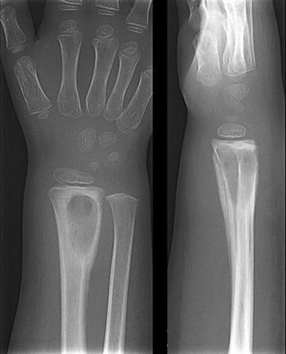
Osteomyelitis refers to infection of the bone, bone marrow with or without extension into the surrounding soft tissues and classically presents with the cardinal signs of inflammation – fever, pain, swelling and loss of mobility. Commonly occurring in children < 5 years of age with nonspecific features (4), it can be challenging to diagnose. The most common site of infection is the metaphysis of the long bones, specifically the femur and the tibia(10).

How does it happen?

The metaphysis is the primary site of infection because of its unique vascular characteristics in the growing skeleton, namely abundant blood supply and capillary lakes or pools where bacteria can lodge and multiply (9, 10). For the first 18 months, there are transphysial vessels from the metaphysis to the epiphysis which traverse the physial plate, translating into a greater risk of spread of infection into the epiphysis and joint space which can result in septic arthritis. After 12 to 18 months, growth of dedicated epiphyseal nutrient vessel in the child and the concomitant disappearance of the transphysial vessels means that the physis can serve as a natural border to prevent the spread of infection from the metaphysis to the epiphysis and joint.

What do I need to know?

The diagnosis of osteomyelitis can portend significant changes to the patient’s management; therefore, it is critical to have answers to these three questions while reviewing imaging to make the most difference in patient care (9).

1. Infection – figure out if there is an infection

2. If yes, then where – localize the infection site

3. If yes, then how – estimate the burden of infection, see if there any drainable collections

Step 1

Radiographs are always obtained first but may not show changes in the early stages except for subtle soft tissue edema. Osteolysis and periosteal reaction are late findings that may take up to 7-10 days to appear, subacute changes include mixed lysis and sclerosis (4, 11). MRI with Gadolinium contrast is the modality of choice for prompt diagnosis of acute osteomyelitis. Any treatment delay can lead to a host of potential complications including septic arthritis, subperiosteal abscess, pyomyositis, deep vein thrombosis, chronic infection, and septicemia with impending death (9). Classically, osteomyelitis presents as decreased T1 and increased T2 signal (due to marrow edema) with enhancement of the affected area on contrast administration and significant surrounding soft tissue edema.

Wrist Radiograph - Osteomyelitis of radius
A 3-year-old boy with osteomyelitis of the distal radius. Anterior-posterior and lateral conventional radiographs show a sharply delineated lucent lesion in the distal radius. Image courtesy: van Schuppen J, van Doorn MM, van Rijn RR. Childhood osteomyelitis: imaging characteristics. Insights Imaging. 2012;3(5):519-533. doi:10.1007/s13244-012-0186-8 (Image credit: van Schuppen J, van Doorn MM, van Rijn RR. Childhood osteomyelitis: imaging characteristics. Insights Imaging. 2012;3(5):519-533. doi:10.1007/s13244-012-0186-8)

Step 2

The next step is to localize the site of infection to quantify disease burden and increase the diagnostic yield of tissue sampling (which is usually not necessary).

Step 3

The final step in the evaluation of osteomyelitis is to establish the presence or absence of drainable fluid collections which may require surgical drainage besides standard surgical debridement of the affected site. Note any intraosseous abscesses –which will appear as an area of high signal intensity in the bone on STIR and T2 weighted images and as a non enhanced center surrounded by rim of enhanced tissue on contrast enhanced images. Note any subperiosteal collections –which will appear as high signal intensity fluid collections trapped between the hypointense linear bony cortex and hypointense lifted off periosteum on T2 weighted and STIR images. On T1 weighted images, high signal intensity globules can sometimes be seen within the abscess, these correspond to fat globules which arise from the bone marrow fat and confirm the subperiosteal nature of the abscess (9).

Imaging series of Hip - Plain Film/US/MRI
A 3-month-old boy with a fever and reluctance to use his right limb. Physical examination showed a swollen, red hip. A. Radiograph shows extensive destruction of the proximal right femur and swollen soft tissue. B. Static ultrasound demonstrates slightly complex predominantly anechoic fluid overlying the eroded right femoral head and neck region. C. Coronal STIR image demonstrates a well-defined complex fluid collection in the right femoral head with central T2 hypointensity representing necrosis. D. Axial T1 image through the bilateral femoroacetabular joints demonstrates complex T1 hypointensity in the right femoral head. E. Diffusion-weighted axial image through the right femoral head and neck demonstrates a restricting fluid collection within the affected femur suggestive of an intraosseous abscess. Source: van Schuppen J, van Doorn MM, van Rijn RR. Childhood osteomyelitis: imaging characteristics. Insights Imaging. 2012;3(5):519-533. doi:10.1007/s13244-012-0186-8 (Image credit: van Schuppen J, van Doorn MM, van Rijn RR. Childhood osteomyelitis: imaging characteristics. Insights Imaging. 2012;3(5):519-533. doi:10.1007/s13244-012-0186-8)

Selected References

4.     Bomer, J. and Holscher, H., 2020. The Radiology Assistant: Hip Pathology In Children. [online] Radiologyassistant.nl. Available at: <https://radiologyassistant.nl/pediatrics/hip/hip-pathology-in-children> [Accessed 8 August 2020].

9.     Jaramillo, D., Dormans, J. P., Delgado, J., Laor, T., & St Geme, J. W. (2017). Hematogenous osteomyelitis in infants and Children: Imaging of a Changing Disease. In Radiology (Vol. 283, Issue 3, pp. 629–643). Radiological Society of North America Inc. https://doi.org/10.1148/radiol.2017151929

10.   van Schuppen, J., van Doorn, M. M. A. C., & van Rijn, R. R. (2012). Childhood osteomyelitis: Imaging characteristics. In Insights into Imaging (Vol. 3, Issue 5, pp. 519–533). Springer. https://doi.org/10.1007/s13244-012-0186-8

11.   Ciambotti, J., Lai, W., Cook, C., Altes, T., Casey, E., Pirttima, T., Pirttima, S., Mentore, K., HIgginbotham, J., Gupta, A., Mcllhenny, J. and Gay, S., 2020. Osteomyelitis. [online] Med Ed Virginia. Available at: < https://www.med-ed.virginia.edu/courses/rad/peds/ms_webpages/ms6bosteomyelitis.html> [Accessed 8 August 2020].

Was this article helpful?
Like 9 Dislike 0
UAMS College of Medicine LogoUAMS College of MedicineUniversity of Arkansas for Medical Sciences
Mailing Address: 4301 West Markham Street, Little Rock, AR 72205
Phone: (501) 686-7000
  • Facebook
  • X
  • Instagram
  • YouTube
  • LinkedIn
  • Pinterest
  • Disclaimer
  • Terms of Use
  • Privacy Statement
  • Legal Notices

© 2026 University of Arkansas for Medical Sciences