• Skip to main content
  • Skip to main content
Choose which site to search.
University of Arkansas for Medical Sciences Logo University of Arkansas for Medical Sciences
College of Medicine: Department of Radiology
  • UAMS Health
  • Jobs
  • Giving
  • About Us
    • News
    • Radiology Alliance for Community & Engagement
    • Giving
    • Contact Us
    • Where We Work
    • Faculty Recruitment
  • Faculty
  • Divisions
    • Neurointerventional Radiology
      • About Us
    • Interventional Radiology
    • Pediatric Radiology
  • Residency Programs
    • Diagnostic Radiology Residency Program
      • Meet Our Residents
      • Your Education
        • Pediatric Radiology On Call Survival Guide
        • Internship Year
        • Boot Camp
        • R1 Education
        • Conferences
        • Clinical Rotations
        • On Call Coverage
        • Mentoring
        • Board Prep
        • Moonlighting
      • Facilities
      • Living in Little Rock
      • Fellowship Placements
      • Wellness
      • Salary and Benefits
    • Interventional Radiology Integrated Residency
      • Guidelines for Selection
      • Clinical Sites
      • Training
      • Faculty
      • Meet Our Residents
      • Program Benefits
    • Interventional Radiology Independent Residency Program
      • Guidelines for Selection
      • Clinical Rotation Sites
      • Block Diagram
      • Training
      • Meet Our Faculty
      • Meet our Resident
      • Benefits
    • Early Specialization in Interventional Radiology Program
    • Newsletter Archive
    • Nuclear Medicine Training Program
    • Application Process
      • Frequently Asked Questions
    • Awards
    • Resident Scrapbook
  • Fellowships
    • Body MRI
    • Breast Imaging
    • Neuroradiology
    • Pediatric Radiology
  • Medical Students
    • Interventional Radiology Interest Group
    • Radiology Interest Group
    • Senior Electives
    • Summer in Radiology
    • The Dr. and Mrs. W.C. Culp Research Student Grant
    • M3s Secure
    • M4s Secure
    • Radiology Technology
  • Research
    • Annual Conferences
    • Ongoing Research
    • Presentations and Posters
    • Recent Publications
    • Resident Research
    • Research Help
    • Radiology Research Symposium
  1. University of Arkansas for Medical Sciences
  2. College of Medicine
  3. Department of Radiology
  4. Ovarian Torsion

Ovarian Torsion

Last modified: October 13, 2022
You are here:
  • KB Home
  • Miscellaneous
  • Ovarian Torsion

What is it?

Torsion of the ovary is defined as partial or complete rotation of the ovary around its axis i.e. vascular stalk causing obstruction of the venous (and lymphatic) outflow and eventually impeding arterial inflow due to increasing edema.

How does it happen?

Typically painful and sudden in onset, ovarian torsion usually presents with severe unilateral pain, commonly on the right side due to greater mobility of the elongated utero-ovarian ligament on the right and the presence of the sigmoid colon on the left (1, 3) which protects against ovarian torsion. Sometimes, patients can present with insidious symptoms due to intermittent torsion/detorsion of the ovary.

Although ectopic pregnancy is an important differential if the patient is pregnant, ovarian torsion still remains high on the differential for the pregnant patient because an early corpus luteum cyst can be the predisposing source of torsion (3). As such, the presence of ovarian cysts (including the differential of cyst rupture) predisposes the patient to ovarian torsion as does a history of prior torsion. Adnexal torsion is twisting of both the ovary and fallopian tube around the vascular pedicle which may be clinically identified as an adnexal mass in the region of maximal abdominal tenderness.

What I need to know?

Like testicular torsion, ovarian torsion is a true gynecologic surgical emergency and early diagnosis can help prevent loss of ovary. Sonographic evaluation for ovarian torsion has high accuracy (74.6%) (2) with inclusion of Doppler interrogation increasing the diagnostic specificity.

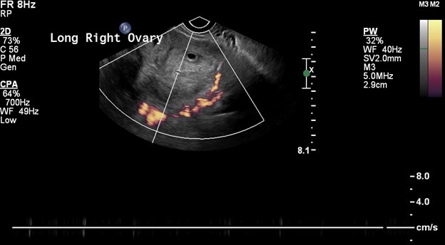
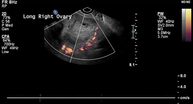
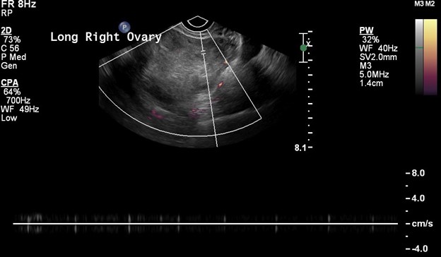
Ultrasound signs to look for while evaluating for ovarian torsion include:

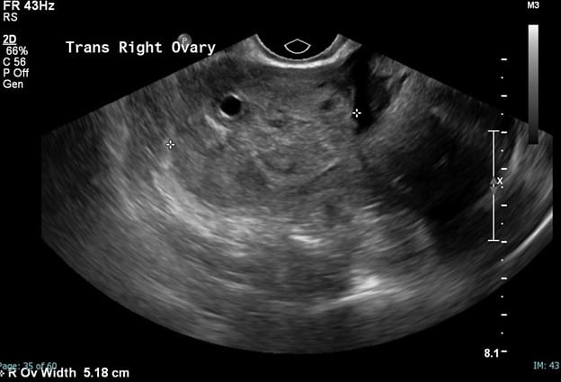
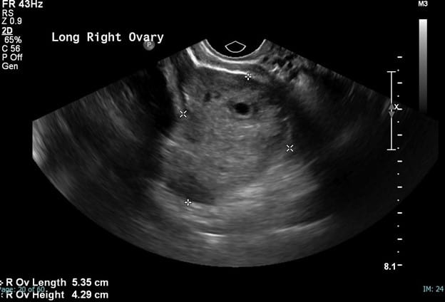
  • Enlarged and edematous ovarian stroma with peripherally displaced follicles – most consistent finding (1))
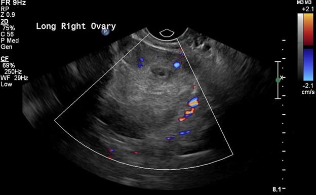
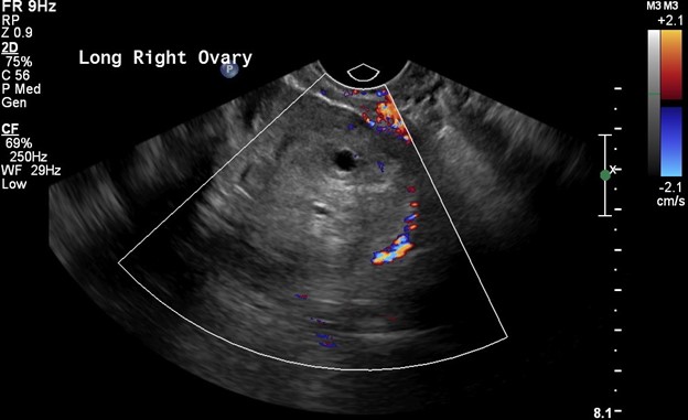
  • Abnormal ovarian vascular flow, initially characterized by lack of central venous flow followed by eventual impairment in arterial flow due to developing edema (note, the ovary has a dual blood supply – ovarian and uterine artery – so do not rule out torsion simply on the basis of preserved arterial flow (1, 3))
  • Associated ovarian mass or cyst predisposing to torsion (usually >5 cm)
  • Whirlpool sign of twisted vascular pedicle and visualized as a snail shell on grayscale imaging (1, 4)

Ovarian Torsion (6 Images):

greyscale US image of the ovary
Transvaginal greyscale ultrasound images of the right ovary demonstrates an enlarged, heterogenous, ill-defined right ovarian tissue with a single dominant follicle.
color US image of the ovary
Color Doppler images demonstrate poor perfusion.
power doppler US image of the ovary
On switching to the power Doppler as seen on this fourth image, no flow is demonstrated within the ovarian parenchyma.
power doppler US image of the ovary
Note absent waveforms on the fourth, fifth, and sixth image. These findings are demonstrative of right ovarian torsion.

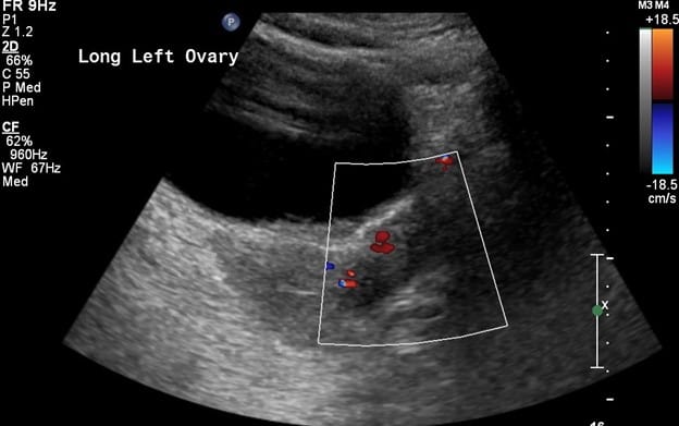
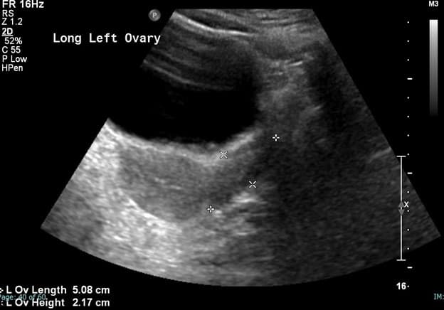
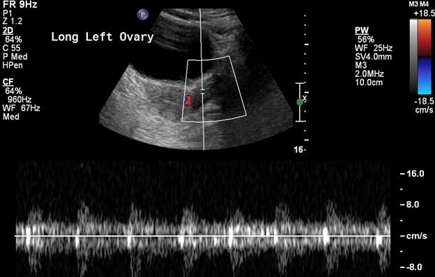
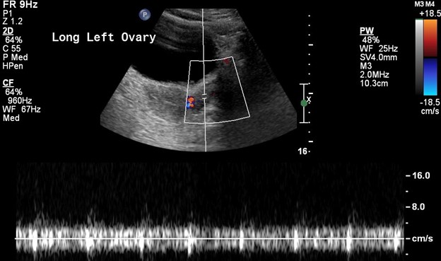
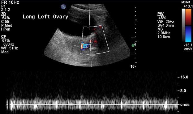
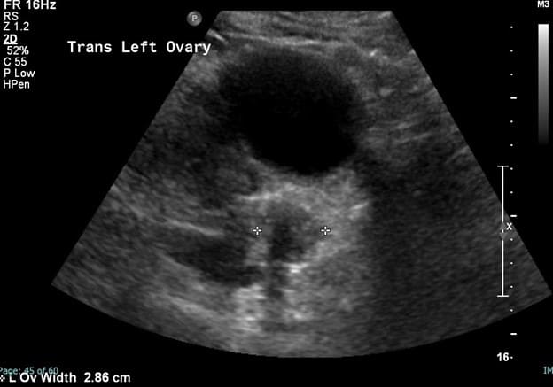
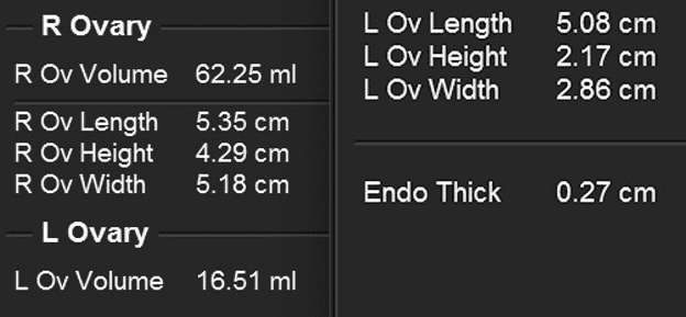
Ovarian Torsion – Compare Ovaries (7 images):

Transabdominal ultrasound images of the left ovary demonstrating well-defined ovarian tissue adjacent to the uterine fundus with preserved color and spectral waveforms.

This last image captures the discrepancy between the ovarian sizes. Note the left ovarian volume which measures normal at 17 mL and the abnormal right ovarian volume which is increased and measures 63 mL.

Selected References:

  1. Govindarajan, K., Nelson, C., Windle, M., & Cendron, M. (2020). Pediatric Testicular Torsion: Background, Anatomy, Pathophysiology. Retrieved 5 September 2020, from https://emedicine.medscape.com/article/2035074-overview#a6
  2. Lonergan, G. (2020). The Radiology Assistant : Acute Scrotum in Children. Retrieved 5 September 2020, from https://radiologyassistant.nl/pediatrics/acute-scrotum/acute-scrotum-in-children
  3. Günther, P., & Rübben, I. (2012). The Acute Scrotum in Childhood and Adolescence. Deutsches Arzteblatt International, 109(25), 449–458. https://doi.org/10.3238/arztebl.2012.0449
  4. Baldisserotto, M., de Souza, J. C. K., Pertence, A. P., & Dora, M. D. (2005). Color Doppler Sonography of Normal and Torsed Testicular Appendages in Children. American Journal of Roentgenology, 184(4), 1287–1292. https://doi.org/10.2214/ajr.184.4.01841287

Was this article helpful?
Like 8 Dislike 0
UAMS College of Medicine LogoUAMS College of MedicineUniversity of Arkansas for Medical Sciences
Mailing Address: 4301 West Markham Street, Little Rock, AR 72205
Phone: (501) 686-7000
  • Facebook
  • X
  • Instagram
  • YouTube
  • LinkedIn
  • Pinterest
  • Disclaimer
  • Terms of Use
  • Privacy Statement
  • Legal Notices

© 2025 University of Arkansas for Medical Sciences