• Skip to main content
  • Skip to main content
Choose which site to search.
University of Arkansas for Medical Sciences Logo University of Arkansas for Medical Sciences
College of Medicine: Department of Radiology
  • UAMS Health
  • Jobs
  • Giving
  • About Us
    • News
    • Radiology Alliance for Community & Engagement
    • Giving
    • Contact Us
    • Where We Work
    • Faculty Recruitment
  • Faculty
  • Divisions
    • Neurointerventional Radiology
      • About Us
    • Interventional Radiology
    • Pediatric Radiology
  • Residency Programs
    • Diagnostic Radiology Residency Program
      • Meet Our Residents
      • Your Education
        • Pediatric Radiology On Call Survival Guide
        • Internship Year
        • Boot Camp
        • R1 Education
        • Conferences
        • Clinical Rotations
        • On Call Coverage
        • Mentoring
        • Board Prep
        • Moonlighting
      • Facilities
      • Living in Little Rock
      • Fellowship Placements
      • Wellness
      • Salary and Benefits
    • Interventional Radiology Integrated Residency
      • Guidelines for Selection
      • Clinical Sites
      • Training
      • Faculty
      • Meet Our Residents
      • Program Benefits
    • Interventional Radiology Independent Residency Program
      • Guidelines for Selection
      • Clinical Rotation Sites
      • Block Diagram
      • Training
      • Meet Our Faculty
      • Meet our Resident
      • Benefits
    • Early Specialization in Interventional Radiology Program
    • Nuclear Medicine Training Program
    • Application Process
      • Frequently Asked Questions
    • Awards
    • Resident Scrapbook
  • Fellowships
    • Body MRI
    • Breast Imaging
    • Neuroradiology
    • Pediatric Radiology
  • Medical Students
    • Interventional Radiology Interest Group
    • Radiology Interest Group
    • Senior Electives
    • Summer in Radiology
    • The Dr. and Mrs. W.C. Culp Research Student Grant
    • M3s Secure
    • M4s Secure
    • Radiology Technology
  • Research
    • Annual Conferences
    • Ongoing Research
    • Presentations and Posters
    • Recent Publications
    • Resident Research
    • Research Help
    • Radiology Research Symposium
  1. University of Arkansas for Medical Sciences
  2. College of Medicine
  3. Department of Radiology
  4. Pyloric Stenosis

Pyloric Stenosis

Last modified: October 8, 2022
You are here:
  • KB Home
  • Gastrointestinal
  • Pyloric Stenosis

What is it?

Hypertrophic pyloric stenosis is the commonest acquired condition with an incidence of about 3 per 1,000 live births per year (10) in otherwise healthy neonates presenting with projectile non-bilious vomiting. Presenting between the sixth through twelfth weeks of life, Caucasian male first-born babies with a family history are the majority of the patient population (outnumbering females by 4 to 1) (2, 10).  

How does it happen?

First there is idiopathic thickening of the circular muscular layer in the pylorus which results in the inability of the pyloric channel to relax and allow for the passage of gastric contents causing gastric outlet obstruction (GOO). It is often said that pyloric stenosis is the only clinical condition that can be felt (palpable “olive”) and seen (as waves of peristalsis traversing the midline) by the clinician (11).  

What do I need to know?

Ultrasound is the ideal diagnostic modality for accurate, non-invasive and rapid confirmation of pyloric stenosis.  

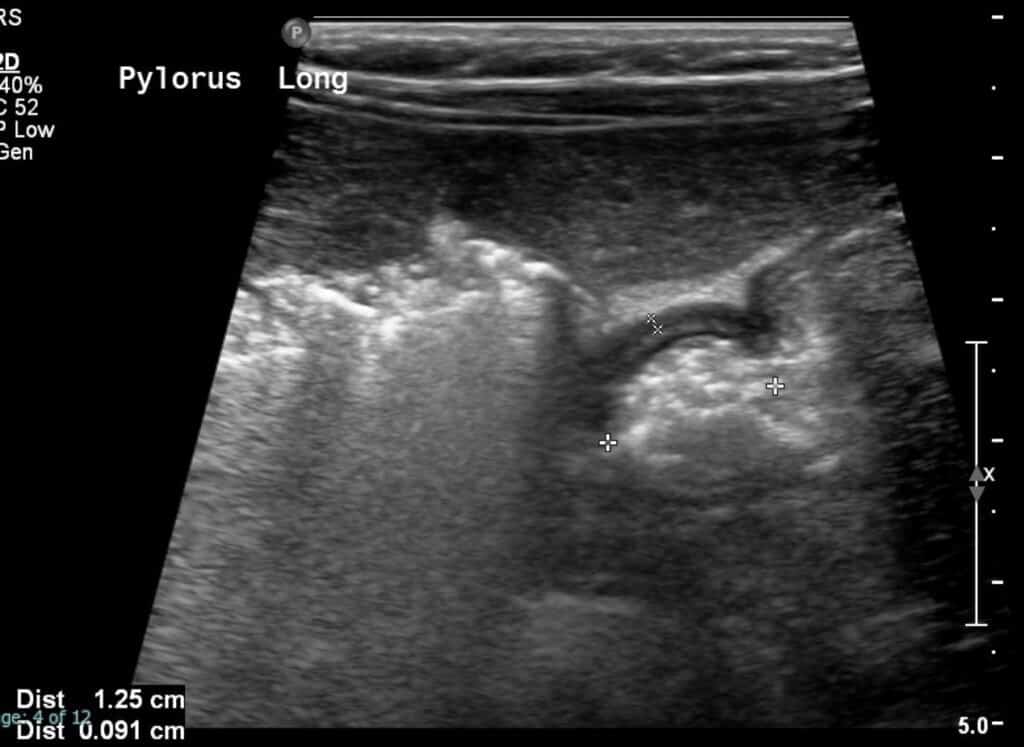
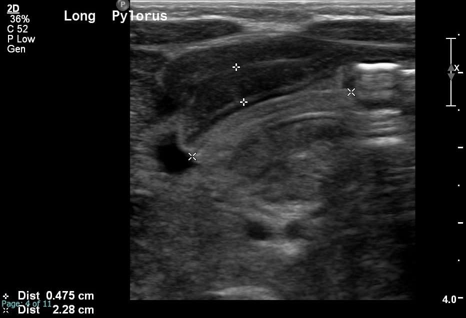
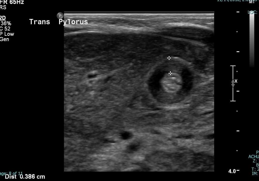
Sonographic findings demonstrate: 

  • Thickened hypoechoic muscular layer >3 mm in size  
    • On sagittal images, the thickened pylorus often resembles the appearance of the uterine cervix 
    • On transverse images, the thickened pylorus has targetoid pattern 
  • No passage of gastric contents through the pyloric canal 
  • Transverse pyloric diameter (wall to wall) >14 mm 
  • Abnormal elongation of the canal >15 mm in length 

These important measurement values can be remembered by the mathematical “pi” sign (3.1415) where 3 represents the thickened muscular layer, 14 represents the transverse pyloric dimension and 15 represents the longitudinal measurement of the pyloric canal (12). 

Static ultrasound grayscale image of the pylorus in its longitudinal dimension demonstrates normal wall thickness.
Cine grayscale images of the open pylorus demonstrate echogenic contents passing through the open pylorus. You may want to play the cine loop several times to appreciate this.

Cine grayscale images of the open pylorus demonstrate echogenic contents passing through the open pylorus. You may want to play the cine loop several times to appreciate this.

Ultrasound image of pylorus
Static grayscale image of pylorus in its longitudinal length demonstrates abnormal thickening of the hypoechoic pyloric muscle which measures up to 0.5 cm (normal < 0.3 cm). Note elongated pyloric channel which measures 2.3 cm (normal < 1.5 cm).
Ultrasound image of pylorus
Static grayscale ultrasound images of the pylorus in its transverse axis demonstrates a thickened hypoechoic muscular layer.
Cine sonographic images of the pylorus demonstrate pyloric canal elongation and thickening ofthe pyloric muscle with echogenic gastric content unable to pass through.

Cine sonographic images of the pylorus demonstrate pyloric canal elongation and thickening of the pyloric muscle with echogenic gastric content unable to pass through.

What do I need to do?

Pay attention to both cine and static images.  The cine images should show echogenic food content unable to pass through the thickened pylorus. Report the dimensions of the pylorus displayed on the static images. Ask the tech to scan down lower to make sure you’re not missing a malrotated bowel if the pylorus looks normal. Basically you should have them scan the upper abdomen transversely in midline so you can look at the SMA/SMV orientation and for the whirlpool sign which may point toward a diagnosis of malrotation. You may want to hop on to read the article on malrotation to refresh your memory if needed.


Selected References

2. S., L. B., Amar, G., & Ellen, P. (2017). Imaging of Pediatric Gastrointestinal Emergencies. Journal of the American Osteopathic College of Radiology, 6(1), 5–14. https://www.jaocr.org/articles/imaging-of-pediatric-gastrointestinal-emergencies

10. Costa Dias, S., Swinson, S., Torrão, H., Gonçalves, L., Kurochka, S., Vaz, C. P., & Mendes, V. (2012). Hypertrophic pyloric stenosis: Tips and tricks for ultrasound diagnosis. In Insights into Imaging (Vol. 3, Issue 3, pp. 247–250). Springer. https://doi.org/10.1007/s13244-012-0168-x

11. Sharwood, E., 2020. Pyloric Stenosis. [online] Don’t Forget The Bubbles. Available at: <https://dontforgetthebubbles.com/pyloric-stenosis/> [Accessed 2 August 2020].

12. Muzio, B. and Qaqish, N., 2020. Hypertrophic Pyloric Stenosis (Ultrasound Measurements Mnemonic) | Radiology Reference Article | Radiopaedia.Org. [online] Radiopaedia.org. Available at: <https://radiopaedia.org/articles/hypertrophic-pyloric-stenosis-ultrasound-measurements-mnemonic-1?lang=us> [Accessed 2 August 2020].

Was this article helpful?
Like 11 Dislike 0
UAMS College of Medicine LogoUAMS College of MedicineUniversity of Arkansas for Medical Sciences
Mailing Address: 4301 West Markham Street, Little Rock, AR 72205
Phone: (501) 686-7000
  • Facebook
  • X
  • Instagram
  • YouTube
  • LinkedIn
  • Pinterest
  • Disclaimer
  • Terms of Use
  • Privacy Statement

© 2025 University of Arkansas for Medical Sciences