Unique Traumatic Fracture Patterns In Kids
What is it?
Acute fractures with different bone break patterns in children compared to adults. Approximately 1 in 3 children are brought to the emergency department after acute trauma (13) due to the generally active lifestyles most adolescents and children have. Plain film radiography remains the mainstay of the imaging armory in keeping with the as low as reasonably achievable (ALARA) principle of ionizing radiation.
How does it happen?
Bones break secondary to moderate to severe acute trauma. Unlike adults, however, children possess growing skeletons which means trauma affects the young child slightly differently when compared to an adult leading to variations in fracture patterns, locations and consequent sequelae of injury. Because the physis is a unique, critical structure in the young skeleton – it is important to be on the lookout for injury to the growth plate not only because it is required to enable future bone growth and any physial disruption can cause devastating deranged growth but also because the cartilaginous physis is weaker, it is more fragile and prone to injury than the ligamentous structures.
What do I need to know?
Look carefully at the physis for any widening. Another common teaching tip is the so-called MARBLE TEST for detecting subtle cortical fractures. Essentially, you should be able to roll a small marble along the cortex of the bone you’re looking at without it bumping into anything or deviating from its smooth path along the bony margins. In this review, we will discuss the Salter Harris classification used for classification of all physial fractures. Long bones are the other common site of trauma in the young child and the distinctive mechanistic properties of the pediatric bone – namely, that it is less stiff and more able to absorb energy before fracturing – make plastic deformation the more typical manifestation than complete bone breaks.
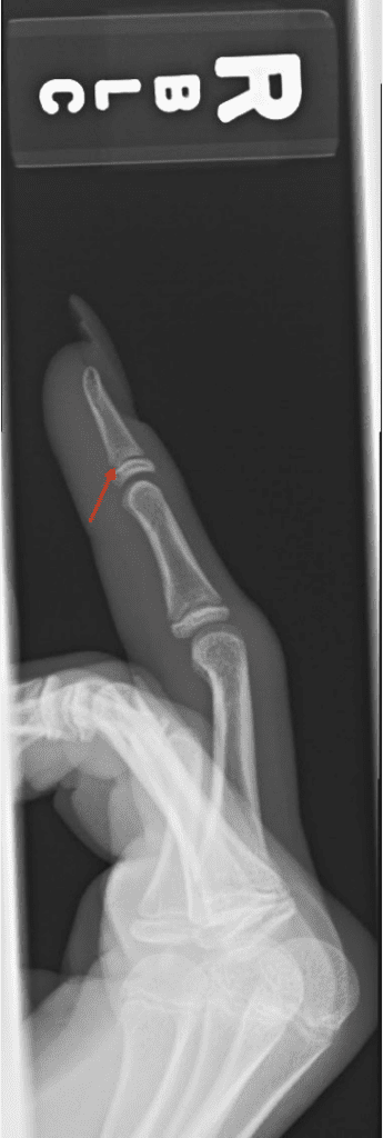
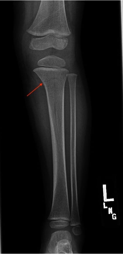
Salter Harris Classification
Note that this classification system is only for injuries involving the growth plate. Type 1 is through the growth plate, separating the metaphysis and epiphysis. Type 2 fractures, the most common type, traverse the physis and the metaphysis. Type 3 fractures in turn, involve the physis and the epiphysis. Type 4 fractures involve all three elements –metaphysis, physis and epiphysis. Type 5 injuries are seen when the growth plate is severely crushed under high axial load stress. This can be remembered by the mnemonic – S A L TE R (14).
S (separated), A (above or away from the joint –involves the growth plate and the structure above the growth plate, i.e. the metaphysis) , L (lower –involves the growth plate and the structure below or lower than the growth plate, i.e. the epiphysis), TE (through everything –all three elements), R (rammed or crushed).
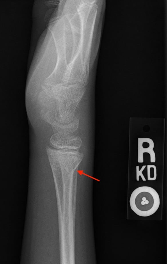
Salter 1 fracture of the left distal radius. Note dorsal translocation of the epiphysis over the growth plate.

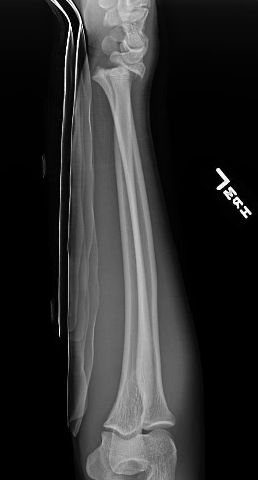
Plastic Deformation
Affected site – forearm, ulna
Features – bone bowing
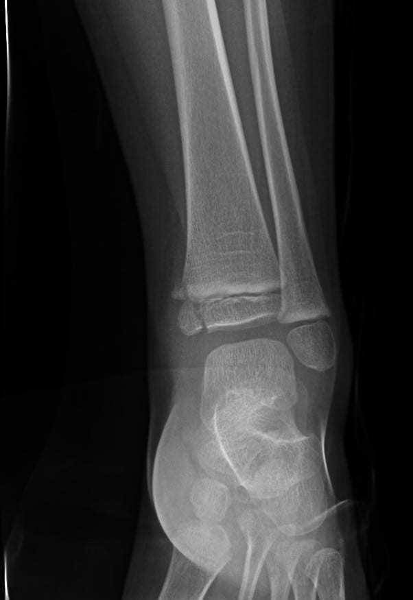
Buckle/Torus fracture
Can be subtle, therefore a high degree of suspicion must be employed.
Affected site –forearm – distal radius/ulna, leg – distal tibia and fibula
Features – small buckle or bump/focal bend in the bony contour
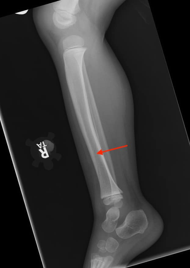
Toddler’s fracture
A type of spiral fracture, toddler’s fracture is commonly encountered in the tibia of children between 9 and 36 months due to new ambulatory stresses placed on the bone. Sometimes confused with trampoline fractures, toddler’s fracture is frequently an occult cause of limping and leg pain in the new walker (16). Classic toddler’s fracture will present as minimally or non-displaced oblique fracture of the middle or distal diaphysis of the tibia (15) with soft tissue swelling at the site.
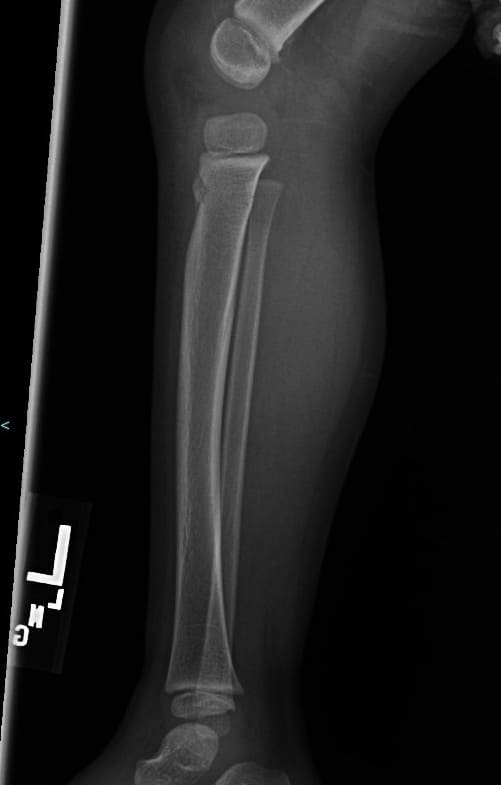
Trampoline fracture
Trampoline fractures, on the other hand, present as transverse hairline fractures through the proximal tibial shaft while jumping on a bouncing trampoline or inflatable bouncy castle. These occur when a second, heavier individual or adult causes the bouncing surface to recoil upwards as the lighter kid (commonly between 2-5 years) is descending downwards leading to a net increase in the combined axial load force (17).
Greenstick fracture
Generally related to FOOSH injuries, these are partial thickness fractures where the bony cortex and periosteum on one side is interrupted while the remainder of the bone is unaffected.
Affected site – can be anywhere, but usually forearm, arm due to fall on outstretched arm
Features – incomplete bending injury with overt fracture on the tension side and plastic deformation on the opposite side of the bone due to compressive forces (18).
Selected References
13. Ho-Fung, V. M., Zapala, M. A., & Lee, E. Y. (2017). Musculoskeletal Traumatic Injuries in Children: Characteristic Imaging Findings and Mimickers. In Radiologic Clinics of North America (Vol. 55, Issue 4, pp. 785–802). W.B. Saunders. https://doi.org/10.1016/j.rcl.2017.02.011
14. Gaillard, F. and Bell, D., 2020. Salter-Harris Fracture Classification (Mnemonic) | Radiology Reference Article | Radiopaedia.Org. [online] Radiopaedia.org. Available at: <https://radiopaedia.org/articles/salter-harris-fracture-classification-mnemonic?lang=us> [Accessed 9 August 2020].
15. Gaillard, F. and Rasuli, B., 2020. Toddler Fracture | Radiology Reference Article | Radiopaedia.Org. [online] Radiopaedia.org. Available at: <https://radiopaedia.org/articles/toddler-fracture?lang=us> [Accessed 9 August 2020].
16. John, S. D., Moorthy, C. S., & Swischuk, L. E. (n.d.). Expanding the Concept of the Toddler’ s Fracture. Radiographics 1997; 17:367-376. Retrieved August 9, 2020, from https://pubs.rsna.org/doi/pdf/10.1148/radiographics.17.2.9084078
17. Dixon, A. and El-Feky, M., 2020. Trampoline Fracture | Radiology Reference Article | Radiopaedia.Org. [online] Radiopaedia.org. Available at: <https://radiopaedia.org/articles/trampoline-fracture?lang=us> [Accessed 9 August 2020].
18. Atanelov Z, Bentley TP. Greenstick Fracture. [Updated 2020 Apr 27]. In: StatPearls [Internet]. Treasure Island (FL): StatPearls Publishing; 2020 Jan-. Available from: https://www.ncbi.nlm.nih.gov/books/NBK513279/