• Skip to main content
  • Skip to main content
Choose which site to search.
University of Arkansas for Medical Sciences Logo University of Arkansas for Medical Sciences
College of Medicine: Department of Radiology
  • UAMS Health
  • Jobs
  • Giving
  • About Us
    • News
    • Radiology Alliance for Community & Engagement
    • Giving
    • Contact Us
    • Where We Work
    • Faculty Recruitment
  • Faculty
  • Divisions
    • Neurointerventional Radiology
      • About Us
    • Interventional Radiology
    • Pediatric Radiology
  • Residency Programs
    • Diagnostic Radiology Residency Program
      • Meet Our Residents
      • Your Education
        • Pediatric Radiology On Call Survival Guide
        • Internship Year
        • Boot Camp
        • R1 Education
        • Conferences
        • Clinical Rotations
        • On Call Coverage
        • Mentoring
        • Board Prep
        • Moonlighting
      • Facilities
      • Living in Little Rock
      • Fellowship Placements
      • Wellness
      • Salary and Benefits
    • Interventional Radiology Integrated Residency
      • Guidelines for Selection
      • Clinical Sites
      • Training
      • Faculty
      • Meet Our Residents
      • Program Benefits
    • Interventional Radiology Independent Residency Program
      • Guidelines for Selection
      • Clinical Rotation Sites
      • Block Diagram
      • Training
      • Meet Our Faculty
      • Meet our Resident
      • Benefits
    • Early Specialization in Interventional Radiology Program
    • Newsletter Archive
    • Nuclear Medicine Training Program
    • Application Process
      • Frequently Asked Questions
    • Awards
    • Resident Scrapbook
  • Fellowships
    • Body MRI
    • Breast Imaging
    • Neuroradiology
    • Pediatric Radiology
  • Medical Students
    • Interventional Radiology Interest Group
    • Radiology Interest Group
    • Senior Electives
    • Summer in Radiology
    • The Dr. and Mrs. W.C. Culp Research Student Grant
    • M3s Secure
    • M4s Secure
    • Radiology Technology
  • Research
    • Annual Conferences
    • Ongoing Research
    • Presentations and Posters
    • Recent Publications
    • Resident Research
    • Research Help
    • Howard J.  Barnhard, MD, Radiology Research Symposium
  1. University of Arkansas for Medical Sciences
  2. College of Medicine
  3. Department of Radiology
  4. Ingested Foreign Body

Ingested Foreign Body

Last modified: October 15, 2022
You are here:
  • KB Home
  • Gastrointestinal
  • Ingested Foreign Body

What is it?

Fairly common in the pediatric population, foreign body ingestion is usually seen in patients 6-36 months (2) as well as in older developmentally delayed children with incidence rates higher in boys and children <1 year of age (2, 18). Most frequently ingested items include coins, toys, jewelry and batteries with button batteries being the most common type (18).

What do I need to know?

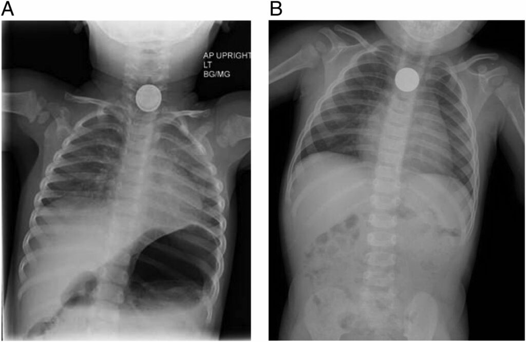
The first step is to differentiate between esophageal ingestion and tracheal aspiration as all airway foreign bodies are medical emergencies. Radiographically, flat foreign bodies orient in a specific plane indicating their location on AP and lateral chest x-rays. Tracheal foreign bodies will orient in the sagittal plane (e.g. –coins will look like flat radiopaque circles on lateral films) while esophageal foreign bodies will orient in the coronal plane (e.g. –coins will look like flat radiopaque circles on AP films). The size, shape, location and number of the foreign bodies should be specifically noted and dictated in the report.

There are 3 main areas in the esophagus where foreign bodies get trapped -thoracic inlet, aortic arch and gastroesophageal junction.

chest X-ray with foreign body
Chest x-ray demonstrates a discoid foreign object in the esophagus with the “halo” or double edge sign suggesting the presence of a button battery (A). Note ingested coin in a different patient (B).  Image courtesy: S. Sarah Green. Ingested and Aspirated Foreign Bodies. Pediatrics in Review Oct 2015, 36 (10) 430-437; DOI: 10.1542/pir.36-10-430. 

Decubitus views are very important to obtain in the case of non-radiopaque foreign bodies.

Although, the majority of ingestions are relatively harmless, there is significant morbidity and mortality caused by the ingestion of button batteries and magnets. Hydroxide radicals produced by the button battery lodged in the esophagus can cause caustic injury, necrosis, perforation, fistula and strictures in the esophagus. Look for the double edge sign to distinguish the button battery from a relatively harmless coin.

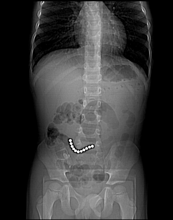
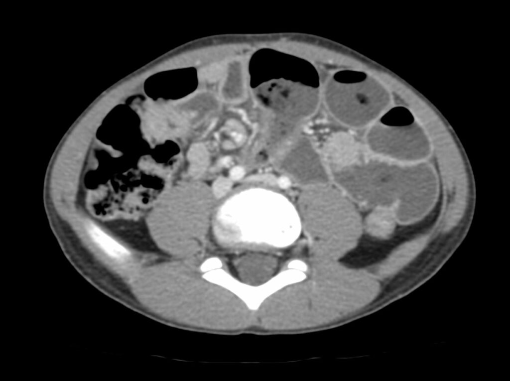
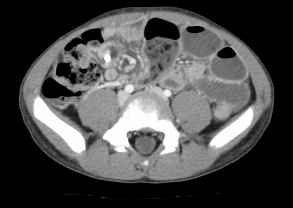
Similarly, ingestion of magnets should raise alarm because when more than one magnet is ingested, magnets will do what magnets know to do – attract each other -leading to a high risk of strong magnetic attraction between ingested magnets as well as pay attention to the magnets through the gastrointestinal walls leading to bowel entrapment which can cause obstruction, necrosis and perforation including entero-enteric fistulas, neatly summarized as “post gastric magnetopathy”.

Scout CT AP localizer radiograph of the abdomen demonstrates a row of circular radiopaque densities within lower abdomen to the right of midline attached to each other suggesting the presence of ingested magnets.
Scout CT AP localizer radiograph of the abdomen demonstrates a row of circular radiopaque densities within lower abdomen to the right of midline attached to each other suggesting the presence of ingested magnets. 
Additional axial post-contrast CT image in the same patient demonstrates a singular partially visualized hyperdense circular foreign object in the right hemiabdomen correlating to known magnets with associated small bowel tethering, ultimately requiring 3 small bowel resections.
This patient unfortunately went on to develop small-bowel obstruction due to bowel entrapment secondary to ingestion of the magnets. Axial postcontrast CT image demonstrates small bowel fecalization sign in the mid abdomen associated with multiple dilated air and fluid-filled loops in the left hemiabdomen. 

Additional axial post-contrast CT image in the same patient demonstrates a singular partially visualized hyperdense circular foreign object in the right hemiabdomen correlating to known magnets with associated small bowel tethering, ultimately requiring 3 small bowel resections.

What do I need to do?

If you see multiple magnets, note the location and number of ingested magnets as well as pay attention to the

  • failure of movement of the magnets through the lumen on sequential radiographs
  • gap between the magnets which can indicate entrapment and raise the possibility of ischemic injury to the intervening bowel (19).

Call the ED up and let them know to talk to surgery.

And to the same effect, if you suspect the child has ingested a button battery instead of an innocuous coin, make sure the ED knows.


Selected References

2. S., L. B., Amar, G., & Ellen, P. (2017). Imaging of Pediatric Gastrointestinal Emergencies. Journal of the American Osteopathic College of Radiology, 6(1), 5–14. https://www.jaocr.org/articles/imaging-of-pediatric-gastrointestinal-emergencies

18. Guelfguat, M., Kaplinskiy, V., Reddy, S. H., & DiPoce, J. (2014). Clinical guidelines for imaging and reporting ingested foreign bodies. In American Journal of Roentgenology (Vol. 203, Issue 1, pp. 37–53). American Roentgen Ray Society. https://doi.org/10.2214/AJR.13.12185

19. Wong, K., Van Tassel, D., Lee, J., Buchmann, R., Riemann, M., Egan, C., & Youssfi, M. (2020). Making the diagnosis of midgut volvulus: Limited abdominal ultrasound has changed our clinical practice. Journal of Pediatric Surgery. https://doi.org/10.1016/j.jpedsurg.2020.04.012

Was this article helpful?
Like 8 Dislike 1
UAMS College of Medicine LogoUAMS College of MedicineUniversity of Arkansas for Medical Sciences
Mailing Address: 4301 West Markham Street, Little Rock, AR 72205
Phone: (501) 686-7000
  • Facebook
  • X
  • Instagram
  • YouTube
  • LinkedIn
  • Pinterest
  • Disclaimer
  • Terms of Use
  • Privacy Statement
  • Legal Notices

© 2026 University of Arkansas for Medical Sciences