• Skip to main content
  • Skip to main content
Choose which site to search.
University of Arkansas for Medical Sciences Logo University of Arkansas for Medical Sciences
College of Medicine: Department of Radiology
  • UAMS Health
  • Jobs
  • Giving
  • About Us
    • News
    • Radiology Alliance for Community & Engagement
    • Giving
    • Contact Us
    • Where We Work
    • Faculty Recruitment
  • Faculty
  • Divisions
    • Neurointerventional Radiology
      • About Us
    • Interventional Radiology
    • Pediatric Radiology
  • Residency Programs
    • Diagnostic Radiology Residency Program
      • Meet Our Residents
      • Your Education
        • Pediatric Radiology On Call Survival Guide
        • Internship Year
        • Boot Camp
        • R1 Education
        • Conferences
        • Clinical Rotations
        • On Call Coverage
        • Mentoring
        • Board Prep
        • Moonlighting
      • Facilities
      • Living in Little Rock
      • Fellowship Placements
      • Wellness
      • Salary and Benefits
    • Interventional Radiology Integrated Residency
      • Guidelines for Selection
      • Clinical Sites
      • Training
      • Faculty
      • Meet Our Residents
      • Program Benefits
    • Interventional Radiology Independent Residency Program
      • Guidelines for Selection
      • Clinical Rotation Sites
      • Block Diagram
      • Training
      • Meet Our Faculty
      • Meet our Resident
      • Benefits
    • Early Specialization in Interventional Radiology Program
    • Newsletter Archive
    • Nuclear Medicine Training Program
    • Application Process
      • Frequently Asked Questions
    • Awards
    • Resident Scrapbook
  • Fellowships
    • Body MRI
    • Breast Imaging
    • Neuroradiology
    • Pediatric Radiology
  • Medical Students
    • Interventional Radiology Interest Group
    • Radiology Interest Group
    • Senior Electives
    • Summer in Radiology
    • The Dr. and Mrs. W.C. Culp Research Student Grant
    • M3s Secure
    • M4s Secure
    • Radiology Technology
  • Research
    • 2026 UAMS Department of Radiology Faculty Research Seed Grant Award
    • Annual Conferences
    • Ongoing Research
    • Presentations and Posters
    • Recent Publications
    • Resident Research
    • Research Help
    • Howard J.  Barnhard, MD, Radiology Research Symposium
  1. University of Arkansas for Medical Sciences
  2. College of Medicine
  3. Department of Radiology
  4. Misplaced Lines And Tubes

Misplaced Lines And Tubes

Last modified: October 13, 2022
You are here:
  • KB Home
  • ICU
  • Misplaced Lines And Tubes

As critically infants often need life saving devices placed to reduce infant mortality and morbidity in the intensive care units, it is important to be familiar with the normal placement of these lines and tubes as a malpositioned line can have dire consequences for the patient. A simple portable chest radiograph is all that is needed to ensure proper tube positioning (1, 3). Here are some pediatric specific lines with a short discussion of their appropriate positioning:

  1. Umbilical artery catheter
    • Use – provides direct access for fast and accurate blood pressure measurement, arterial blood gas sampling and IV access for both fluids and medications in babies <5 days old
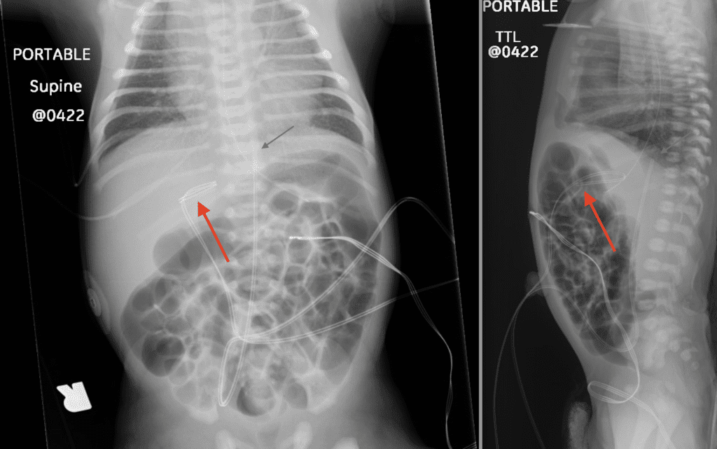
    • Placement – Passes through the patent umbilical artery and makes a U turn to enter the internal iliac artery before entering the aorta, lies posteriorly on lateral radiographs
    • Appropriate Position – avoid carotid and renal origins, can be in
      High position: T6-10
      Low position: L3-L5 (1, 2)
  2. Umbilical Venous Line
    • Use – provides direct access for parenteral nutrition, venous blood sampling and IV access for both fluids and medications in babies <5 days old
    • Placement – Passes through the patent umbilical vein into the left portal vein, ductus venosus and into the IVC through the hepatic vein, lies anteriorly on lateral radiographs
    • Appropriate Position – At the level of T8-9 (1)
      • Avoid low position in the umbilical vein, or in the portal venous system (can cause thrombosis)
      • Avoid high position in the right (or left through patent foramen ovale or atrial septal defect) atrium (can cause arrhythmia or perforation) (2)
Frontal and Cross Table X-Rays - Umbilical Lines
Frontal and cross table lateral views of the chest and abdomen in this neonate demonstrate multiple umbilical lines. Red arrow points to the malpositioned UVC catheter which is seen terminating in the portal venous system. Green arrow points to the high position (T10) of the UAC catheter (appropriate positioning).

For more leisurely reading, check out this article on Radiology Assistant to learn about all the NICU lines and tubes.


Selected References

  1. Jain, S. N. (2011). A pictorial essay: Radiology of lines and tubes in the intensive care unit. In Indian Journal of Radiology and Imaging (Vol. 21, Issue 3, pp. 182–190). Wolters Kluwer — Medknow Publications. https://doi.org/10.4103/0971-3026.8536
  2. van Schuppen, J., Onland, W., & van Rijn, R. (2020). The Radiology Assistant : Lines and tubes in Neonates. Retrieved 13 September 2020, from https://radiologyassistant.nl/pediatrics/lines-and-tubes/lines-and-tubes-in-neonates
  3. Aquino SL. Routine chest radiograph. ACR appropriateness criteria, 2006. American College of Radiology. [Last accessed on 2020 Sep 12]. Available from: http://www.acr.org.

Was this article helpful?
Like 2 Dislike 0
UAMS College of Medicine LogoUAMS College of MedicineUniversity of Arkansas for Medical Sciences
Mailing Address: 4301 West Markham Street, Little Rock, AR 72205
Phone: (501) 686-7000
  • Facebook
  • X
  • Instagram
  • YouTube
  • LinkedIn
  • Pinterest
  • Disclaimer
  • Terms of Use
  • Privacy Statement
  • Legal Notices

© 2026 University of Arkansas for Medical Sciences