• Skip to main content
  • Skip to main content
Choose which site to search.
University of Arkansas for Medical Sciences Logo University of Arkansas for Medical Sciences
College of Medicine: Department of Microbiology and Immunology
  • UAMS Health
  • Jobs
  • Giving
  • About
    • Campus, City, and State
    • Department News
    • Maps & Directions
  • Faculty
    • Primary Faculty
      • Jon Blevins, Ph.D.
      • Youssef Aachoui, Ph.D.
      • Karl Boehme, Ph.D.
      • Martin Cannon, Ph.D.
      • Mohamed O. Elasri, Ph.D.
      • Craig Forrest, Ph.D.
      • Lu Huang, Ph.D.
      • Clay Jackson-Litteken, Ph.D.
      • Lin-Xi Li, Ph.D.
      • Matthew Jorgenson, Ph.D.
      • Chia Lee, Ph.D.
      • Jia Liu, Ph.D.
      • Mark Manzano, Ph.D.
        • Manzano Lab
          • Team
          • Publications
          • News
          • Virology Lab Meeting (Fall ’25)
          • Join Us
      • Mark Smeltzer, Ph.D.
      • Jason Stumhofer, Ph.D.
      • Daniel E. Voth, Ph.D.
        • Voth Lab
      • Tiffany Weinkopff, Ph.D.
      • Xuming Zhang, Ph.D.
  • Staff
    • Administrative Staff
    • Postdoctoral Fellows
      • Marena Guzman, Ph.D.
      • Changhoon Oh, Ph.D.
      • Shana Owens, Ph.D.
    • Research Staff
  • Graduate Program
    • Ph.D. & M.D./Ph.D. Rotations in MBIM Track
    • Courses Offered
    • Graduate Students
  • Research Cores
    • Center for Microbial Pathogenesis and Host Inflammatory Responses
      • About Us
        • COBRE-Related Publications
        • Contact Us
        • Mission Statement
      • Advisory Boards
        • External
        • Internal
      • Cores
        • Administrative Resources
        • Technical
      • Research
        • Pilot Projects
        • Project Leaders
        • Start-up Projects
    • DNA Sequencing Core Facility
      • Preparing Your Samples for Submission (Sanger)
      • Submitting Samples (Sanger)
      • Recommendations, Downloads, and Useful Links (Sanger)
      • TapeStation 4150
      • NextSeq 2000 (Illumina)
      • Covaris ME220
    • Flow Cytometry Core Facility
      • Training and Education
      • Protocols and Reagents
        • Immunofluorescence Staining for Immunotyping
        • Cell Cycle Staining using PI
        • Sorting and Staining Buffers
        • Lysing red blood cells
      • Sample Submission
        • Appointments and Pricing
        • Sample Preparation
      • Instruments
        • BD FACS Symphony- 16 Color Self Service Cytometer
        • BD LSRFortessa- 4 laser 16 color cytometer
        • Cytek Aurora CS-cell sorter
        • BD FACS Celesta-12 color self-service cytometer
        • Cytek Northern Lights -Full Spectrum Cytometer
        • Hematology Analyzer-VETSCAN HM5
      • Data Analysis and Citation
    • Next Generation Sequencing
      • Small Genome Sequencing
      • 16S Metagenomics
      • RNA-Seq
    • iLab-External
    • iLab-UAMS
  • Resources
    • Arkansas Tick-Borne Pathogen Surveillance Program
    • Microbiology & Immunology Research Seminar
  1. University of Arkansas for Medical Sciences
  2. College of Medicine
  3. Department of Microbiology and Immunology
  4. Faculty
  5. Primary Faculty
  6. Matthew Jorgenson, Ph.D.

Matthew Jorgenson, Ph.D.

Matthew Jorgenson, Ph.D.

Assistant Professor

Research Interest: Manipulating the glycans that surround and protect bacteria
Ph.D.: The University of Iowa
Postdoctoral: University of Arkansas for Medical Sciences
Lab: Biomed I, Room 511
Phone: 501-686-7706
Fax: 501-686-5359
Email: MAJorgenson@uams.edu

Currently accepting new students

Current Lab Members

Joseph Bryant, M.S. (Research Associate III)

Emily Robbs, M.S. (Ph.D. Student)

Jorgenson Lab, Fall 2022

Research Description

The Jorgenson lab is focused on investigating the bacterial surface and finding new ways to exploit its properties.

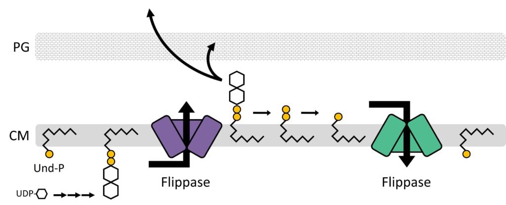
How do bacteria coat themselves?  Bacteria surround themselves in sugary layers that protect against environmental hazards, antibiotics, and immune systems. Though formidable, these protective layers harbor a critical weakness: their fates are tied to a vulnerable lipid carrier known as undecaprenyl phosphate (Und-P). We are currently focused on (i) understanding how Und-P is distributed between competing pathways, (ii) defining pathways that are connected to Und-P metabolism, and (iii) exploiting Und-P production for useful purposes. By developing a deeper understanding of Und-P, the more rational and sophisticated our antibacterial therapies will become.

Model for Und-P utilization
Und-P scaffolds bacterial glycan assembly. Phosphosugars are transferred onto the lipid carrier undecaprenyl phosphate (Und-P) to form Und-PP-linked intermediates. After assembly, flippases transfer Und-PP-linked intermediates across the cytoplasmic membrane (CM). Later, glycans are incorporated into surface layers like the peptidoglycan (PG) cell wall, thus releasing Und-PP to be recycled for another round of assembly.

How are bacteria shaped?  Bacteria are encased by a rigid superstructure known as the peptidoglycan cell wall. The cell wall confers shape and is essential to protect bacteria against osmotically induced lysis. While antibiotics target nearly every stage in cell wall assembly, the emergence of antibiotic resistance means that new targets are needed. We are currently focused on characterizing several new morphological pathways that are required to maintain cell wall integrity, as well as searching for more.

How do we address these questions?  To determine how bacteria assemble and maintain their protective layers, we employ genetic, microscopic, and biochemical tools in the model organism Escherichia coli.

Publications

  • Kay EJ, Dooda MK, Bryant JC, Reid AJ, Wren BW, Troutman JM and Jorgenson MA. 2024. Engineering Escherichia coli for increased Und-P availability leads to material improvements in glycan expression technology. Microbial Cell Factories 23:72.
  • Jorgenson MA and Bryant JC. 2021. A genetic screen to identify factors affected by undecaprenyl phosphate recycling uncovers novel connections to morphogenesis in Escherichia coli. Molecular Microbiology 115(2):191-207. PMID: 32979869.
  • Jorgenson MA, MacCain WJ, Meberg BM, Kannan S, Bryant JC, Young KD. 2019. Simultaneously inhibiting undecaprenyl phosphate production and peptidoglycan synthases promotes rapid lysis in Escherichia coli. Molecular Microbiology 112(1):233-248. PMCID: PMC6616000.
  • Jorgenson MA, Young KD. 2018. YtfB, an OapA domain-containing protein, is a new cell division protein in Escherichia coli. Journal of Bacteriology 200(13). PMCID: PMC5996693.
  • Jorgenson MA, Young KD. 2016. Interrupting biosynthesis of O antigen or the lipopolysaccharide core produces morphological defects in Escherichia coli by sequestering undecaprenyl phosphate. Journal of Bacteriology 198 (22):3070-3079. PMCID: PMC5075036.
  • Jorgenson MA, Kannan S, Laubacher ME, Young KD. 2016. Dead-end intermediates in the enterobacterial common antigen pathway induce morphological defects in Escherichia coli by competing for undecaprenyl phosphate. Molecular Microbiology 100(1):1-14. PMCID: PMC4845916.
  • Yahashiri A, Jorgenson MA, Weiss DS. 2015. Bacterial SPOR domain are recruited to septal peptidoglycan by binding to glycan strands that lack stem peptides. Proceedings of the National Academy of Sciences USA 112:11347-11352. 
  • Jorgenson MA, Chen Y, Yahashiri A, Popham DL, Weiss DS. 2014. The bacterial septal ring protein RlpA is a lytic transglycosylase that contributes to rod shape and daughter cell separation in Pseudomonas aeruginosa. Molecular Microbiology 93(1): 113-128. PMCID: PMC4086221.

Dr. Jorgenson’s publications at PubMed.

UAMS College of Medicine LogoUAMS College of MedicineUniversity of Arkansas for Medical Sciences
Mailing Address: 4301 West Markham Street, Little Rock, AR 72205
Phone: (501) 686-7000
  • Facebook
  • X
  • Instagram
  • YouTube
  • LinkedIn
  • Pinterest
  • Disclaimer
  • Terms of Use
  • Privacy Statement
  • Legal Notices

© 2025 University of Arkansas for Medical Sciences